Electrospun Cellulose Fibres and Applications Wan... · 2019-08-29 · dan keterbaharuan....
14
Sains Malaysiana 48(7)(2019): 1459–1472 http://dx.doi.org/10.17576/jsm-2019-4807-15 Electrospun Cellulose Fibres and Applications (Serabut dan Aplikasi Selulosa Elektropusing) WAN FARAHHANIM WAN FATHILAH & RIZAFIZAH OTHAMAN* ABSTRACT Cellulose fibres and nanofibres have gained interest because of the high strength and firmness, biodegradability and renewability. The enthusiasm in cellulose and its modification as cellulose-derivative has been exponentially expanding. This paper discuss on cellulose and its derivatives, and methods to produce cellulose fibres and nanofibres. Emphasis is given on electrospinning technique, the most utilised technique to produce cellulose fibres and cellulose nanofibres with ranging from nanometer to millimeter in diameter. It also summarises cellulose in terms of a matrix of cellulose, solvent, parameter electrospinning, fibre diameter and their perspective applications. Keywords: Cellulose; cellulose nanofibre; electrospinning; electrospun ABSTRAK Serabut selulosa dan serabut nano mendapat perhatian kerana kekuatan yang tinggi dan keteguhan, keterbiodegradan dan keterbaharuan. Keghairahan dalam selulosa dan pengubahsuaiannya sebagai selulosa-terbitan telah berkembang pesat. Kertas ini membincangkan tentang selulosa dan terbitannya serta kaedah untuk menghasilkan serabut selulosa dan serabut nano. Penekanan diberikan pada teknik elektropemusingan, teknik yang paling kerap digunakan untuk menghasilkan serabut selulosa dan selulosa serabut nano dengan diameter antara nanometer hingga milimeter. Ia juga merumuskan selulosa daripada segi matriks selulosa, pelarut, parameter elektropemusingan, diameter serabut dan perspektif aplikasinya. Kata kunci: Elektropemusingan; elektropusing; selulosa; selulosa serabut nano INTRODUCTION As a renewable bio-based material on the earth, cellulose has become an extensive interest in producing novel polymers and materials (Prasanth et al. 2015; Rosenau et al. 2006; Young-Mook Lim 2010). Cellulose with biodegradable properties makes its fibre to be utilised in many applications such as biomedical areas, protective clothing, mechanical areas and filtration (Cucolo & Aminuddin 2001; Muhammad Johan Iskandar et al. 2018; Razali et al. 2018). This homopolysaccharide is produced through β(1–4) glycosidic bonds condensed from linearly connecting D-glucose units as shown in Figure 1. Researchers believed that intermolecular hydrogen bonds in cellulose chain can give the linear polymer molecules to unite in sheet-like structures while intramolecular hydrogen bonds bring cellulose chain stiffness character (Klemm et al. 2005). Nonetheless, dissolution of cellulose has become a challenging issue due to its properties of limited solubility and incapability to melt in common aqueous and organic solvents (Fauzee & Othaman 2013; Krssig 1993; Marsh & Wood 1942). This is caused by the numerous intermolecular and intramolecular hydrogen bond networks exist in cellulose structure (Zhou & Zhang 2000). Besides, other interactions involved among cellulose structures have been mostly omitted. Currently, this complication has been already discussed and reanalysed as an accepted description (Medronho et al. 2012). It has been decided that biopolymeric cellulose network has a clear amphiphilic character and that cellulose solubility has the highest impact of hydrophobic interactions in the cellulose structure (Medronho & Lindman 2015). Thus, it is crucial to promote ‘green’ cellulose extraction technique and suitable cellulose dissolution path to make full use of cellulose resources in the world. The manufactures of polymer nanofibres via electrospinning technique have been perceived as a persuasive system for producing fibres with submicron diameters by electrostatic forces (Bognitzki et al. 2001; Muhammad Hariz et al. 2014; Razali et al. 2018). Many polymers have been effectively electrospun into ultrafine filaments generally in dissolvable arrangement and some in melt structure (Faten Ermala et al. 2017; Huang et al. 2003; Hutmacher & Dalton 2011). Electrospinning is used for producing nanofibres when colloidal suspension of solid nanoparticles or solution is spun (Jaworek et al. 2009; Leach et al. 2011). The reason for choosing electrospinning is that it requires a single step, low energy and low-cost material processing technology that can operate in atmospheric conditions (Yu 2007). Electric charge draws liquid from a nozzle in fine jet and disperses it into highly charged droplets. The free charge generates electric stress, which make the solution to fly over from
Transcript of Electrospun Cellulose Fibres and Applications Wan... · 2019-08-29 · dan keterbaharuan....
Sains Malaysiana 48(7)(2019): 1459–1472 http://dx.doi.org/10.17576/jsm-2019-4807-15
Electrospun Cellulose Fibres and Applications (Serabut dan Aplikasi Selulosa Elektropusing)
WAN FARAHHANIM WAN FATHILAH & RIZAFIZAH OTHAMAN*
ABSTRACT
Cellulose fibres and nanofibres have gained interest because of the high strength and firmness, biodegradability and renewability. The enthusiasm in cellulose and its modification as cellulose-derivative has been exponentially expanding. This paper discuss on cellulose and its derivatives, and methods to produce cellulose fibres and nanofibres. Emphasis is given on electrospinning technique, the most utilised technique to produce cellulose fibres and cellulose nanofibres with ranging from nanometer to millimeter in diameter. It also summarises cellulose in terms of a matrix of cellulose, solvent, parameter electrospinning, fibre diameter and their perspective applications.
Keywords: Cellulose; cellulose nanofibre; electrospinning; electrospun
ABSTRAK
Serabut selulosa dan serabut nano mendapat perhatian kerana kekuatan yang tinggi dan keteguhan, keterbiodegradan dan keterbaharuan. Keghairahan dalam selulosa dan pengubahsuaiannya sebagai selulosa-terbitan telah berkembang pesat. Kertas ini membincangkan tentang selulosa dan terbitannya serta kaedah untuk menghasilkan serabut selulosa dan serabut nano. Penekanan diberikan pada teknik elektropemusingan, teknik yang paling kerap digunakan untuk menghasilkan serabut selulosa dan selulosa serabut nano dengan diameter antara nanometer hingga milimeter. Ia juga merumuskan selulosa daripada segi matriks selulosa, pelarut, parameter elektropemusingan, diameter serabut dan perspektif aplikasinya.
Kata kunci: Elektropemusingan; elektropusing; selulosa; selulosa serabut nano
INTRODUCTION
As a renewable bio-based material on the earth, cellulose has become an extensive interest in producing novel polymers and materials (Prasanth et al. 2015; Rosenau et al. 2006; Young-Mook Lim 2010). Cellulose with biodegradable properties makes its fibre to be utilised in many applications such as biomedical areas, protective clothing, mechanical areas and filtration (Cucolo & Aminuddin 2001; Muhammad Johan Iskandar et al. 2018; Razali et al. 2018). This homopolysaccharide is produced through β(1–4) glycosidic bonds condensed from linearly connecting D-glucose units as shown in Figure 1. Researchers believed that intermolecular hydrogen bonds in cellulose chain can give the linear polymer molecules to unite in sheet-like structures while intramolecular hydrogen bonds bring cellulose chain stiffness character (Klemm et al. 2005). Nonetheless, dissolution of cellulose has become a challenging issue due to its properties of limited solubility and incapability to melt in common aqueous and organic solvents (Fauzee & Othaman 2013; Krassig 1993; Marsh & Wood 1942). This is caused by the numerous intermolecular and intramolecular hydrogen bond networks exist in cellulose structure (Zhou & Zhang 2000). Besides, other interactions involved among cellulose structures have been mostly omitted. Currently, this complication has been already discussed and reanalysed
as an accepted description (Medronho et al. 2012). It has been decided that biopolymeric cellulose network has a clear amphiphilic character and that cellulose solubility has the highest impact of hydrophobic interactions in the cellulose structure (Medronho & Lindman 2015). Thus, it is crucial to promote ‘green’ cellulose extraction technique and suitable cellulose dissolution path to make full use of cellulose resources in the world. The manufactures of polymer nanofibres via electrospinning technique have been perceived as a persuasive system for producing fibres with submicron diameters by electrostatic forces (Bognitzki et al. 2001; Muhammad Hariz et al. 2014; Razali et al. 2018). Many polymers have been effectively electrospun into ultrafine filaments generally in dissolvable arrangement and some in melt structure (Faten Ermala et al. 2017; Huang et al. 2003; Hutmacher & Dalton 2011). Electrospinning is used for producing nanofibres when colloidal suspension of solid nanoparticles or solution is spun (Jaworek et al. 2009; Leach et al. 2011). The reason for choosing electrospinning is that it requires a single step, low energy and low-cost material processing technology that can operate in atmospheric conditions (Yu 2007). Electric charge draws liquid from a nozzle in fine jet and disperses it into highly charged droplets. The free charge generates electric stress, which make the solution to fly over from
1460
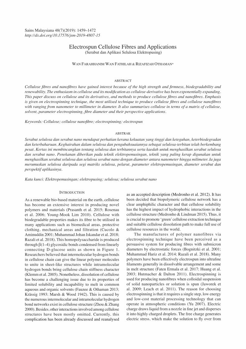
the needle and form a conical shape commonly known as the Taylor cone. The injection of liquid can be controlled by adjusting the flow rate, voltage applied to nozzle, speed of drum controller, distance between tips of needle to drum controller and diameter of the syringe (Javadian et al. 2012; Quan et al. 2010; Zeng et al. 2003). There is a minimum viscosity for every polymer solution termed the critical entanglement concentration, beneath which a steady fly cannot be accomplished and no nanofibre will be framed in spite of the fact that particles may be accomplished, which are beads from electrospraying method (Leach et al. 2011; Reneker & Yarin 2008; Wang et al. 2009). The principle process of electrospinning as shown in Figure 2. This paper attempts to assess the background of cellulose fibres and cellulose nanofibres followed by electrospun cellulose fibres and nanofibres and lastly the applications of cellulose fibres and nanofibres. To date, most of the review papers on cellulose electrospinning either focusing on derivatives or the effect of the electrospinning parameter on the characteristic of the nanofiber (Ohkawa 2015; Prasanth et al. 2015; Young-Mook 2010). The objective of this current study was to discuss on cellulose and its derivatives and how to produce cellulose fibres and nanofibres. However, emphasis is given on electrospinning technique with their perspective applications.
CELLULOSE
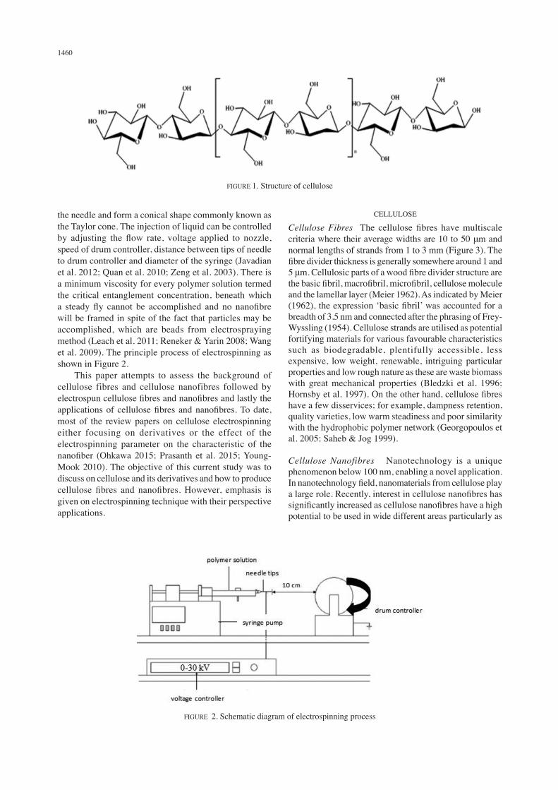
Cellulose Fibres The cellulose fibres have multiscale criteria where their average widths are 10 to 50 μm and normal lengths of strands from 1 to 3 mm (Figure 3). The fibre divider thickness is generally somewhere around 1 and 5 μm. Cellulosic parts of a wood fibre divider structure are the basic fibril, macrofibril, microfibril, cellulose molecule and the lamellar layer (Meier 1962). As indicated by Meier (1962), the expression ‘basic fibril’ was accounted for a breadth of 3.5 nm and connected after the phrasing of Frey-Wyssling (1954). Cellulose strands are utilised as potential fortifying materials for various favourable characteristics such as biodegradable, plentifully accessible, less expensive, low weight, renewable, intriguing particular properties and low rough nature as these are waste biomass with great mechanical properties (Bledzki et al. 1996; Hornsby et al. 1997). On the other hand, cellulose fibres have a few disservices; for example, dampness retention, quality varieties, low warm steadiness and poor similarity with the hydrophobic polymer network (Georgopoulos et al. 2005; Saheb & Jog 1999).
Cellulose Nanofibres Nanotechnology is a unique phenomenon below 100 nm, enabling a novel application. In nanotechnology field, nanomaterials from cellulose play a large role. Recently, interest in cellulose nanofibres has significantly increased as cellulose nanofibres have a high potential to be used in wide different areas particularly as
FIGURE 1. Structure of cellulose
FIGURE 2. Schematic diagram of electrospinning process
1461
the reinforcement in evolution of nanocomposites. Many studies have been done on isolation and characterisation of cellulose nanofibres from various sources (Saurabh et al. 2016). Extraction of cellulose nanofibres can be obtained from the cell walls by simple mechanical methods or a combination of mechanical and chemical methods.
PRODUCTION OF CELLULOSE NANOFIBRES
Chemomechanical Treatment To obtain purified cellulose, Alemdar and Sain (2008a) used wheat straw by a chemical treatment to extract cellulose nanofibres. A mechanical treatment was applied to the chemically treated fibres to diagnose the nanofibres from the cell walls. By a chemomechanical technique, cellulose nanofibres were extracted from the agricultural residues, wheat straw and soy hulls (Alemdar & Sain 2008b). Moreover, the diameters and lengths of wheat straw nanofibres have been determined ranging from 10-80 nm and a few thousand nm, respectively. In a proportion of soy hull nanofibres, the diameter is 20-120 nm with a length shorter than that of wheat straw nanofibres. By applying chemomechanical treatments, Wang and Sain (2007) extracted the cellulose nanofibres from soybean stock. These are an assortment of cellulose nanofibres with 50-100 nm of diameter range and thousands nm in length. After that, wheat straw was extracted to obtain the cellulose nanofibrils using steam explosion, acidic treatment and high shear mechanical treatment.
Homogenisation Process At numerous desirable lengths and diameters below 100 nm from particular starting cellulose materials, Zimmermann et al. (2010) tried to separate nanofibrillated cellulose (NFC) by mechanical diffusion and high pressure (up to 1500 bar) of homogenisation processes. The treatment eventuated in nanoscaled fibril networks. Two profitable fibrous celluloses produced bigger cellulose aggregates with micrometer dimensions and less homogeneous complex structure. Besides that, alkaline-treated pulp was soaked
overnight in an 8 wt. % solution of H2O2 (v/v). Then, bleached pulp was rinsed with abundant distilled water. Bleached pulp was next treated with 10% HCl (1 N) solution and mixed at temperature around 60 ± 1°C for 5 h using ultrasonicator. Finally, to neutralize the final pH, the fibres were taken out and washed a few times with distilled water and then dried. Fibres were suspended in water and continuously stirred for 15 min by practicing high shear homogeniser. High-shearing action resolves the fibre agglomerates and resulted in nanofibrils (Kaushik et al. 2010). Moreover, cellulose nanofibres have been developed by dissolving cellulose in solvents including ethylene diamine with its salt selected from the group consisting potassium thiocyanate and potassium iodide where the salt is present at their saturation points (Frey & Yong 2004).
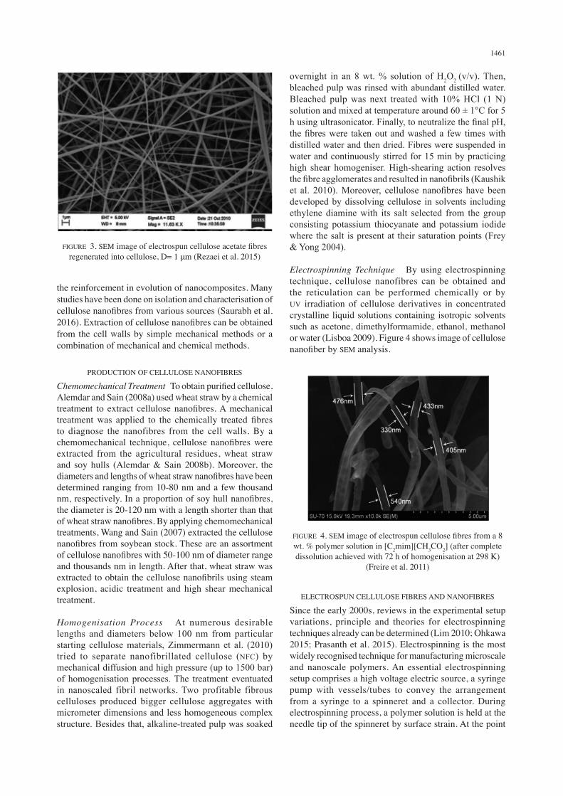
Electrospinning Technique By using electrospinning technique, cellulose nanofibres can be obtained and the reticulation can be performed chemically or by UV irradiation of cellulose derivatives in concentrated crystalline liquid solutions containing isotropic solvents such as acetone, dimethylformamide, ethanol, methanol or water (Lisboa 2009). Figure 4 shows image of cellulose nanofiber by SEM analysis.
FIGURE 3. SEM image of electrospun cellulose acetate fibres regenerated into cellulose, D= 1 μm (Rezaei et al. 2015)
FIGURE 4. SEM image of electrospun cellulose fibres from a 8 wt. % polymer solution in [C2mim][CH3CO2] (after complete dissolution achieved with 72 h of homogenisation at 298 K)
(Freire et al. 2011)
ELECTROSPUN CELLULOSE FIBRES AND NANOFIBRES
Since the early 2000s, reviews in the experimental setup variations, principle and theories for electrospinning techniques already can be determined (Lim 2010; Ohkawa 2015; Prasanth et al. 2015). Electrospinning is the most widely recognised technique for manufacturing microscale and nanoscale polymers. An essential electrospinning setup comprises a high voltage electric source, a syringe pump with vessels/tubes to convey the arrangement from a syringe to a spinneret and a collector. During electrospinning process, a polymer solution is held at the needle tip of the spinneret by surface strain. At the point
1462
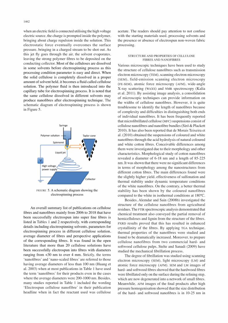
when an electric field is connected utilising the high voltage electric source, the charge is prompted inside the polymer, bringing about charge repulsion inside the solution. This electrostatic force eventually overcomes the surface pressure, bringing in a charged stream to be shot out. As this jet fly goes through the air, the solvent evaporates, leaving the strong polymer fibres to be deposited on the conducting collector. Most of the celluloses are dissolved in some solvents before electrospinning process as this processing condition parameter is easy and direct. When the solid cellulose is completely dissolved in a proper amount of solvent held, it becomes a fluid called cellulose solution. The polymer fluid is then introduced into the capillary tube for electrospinning process. It is noted that the same cellulose dissolved in different solvents may produce nanofibres after electrospinning technique. The schematic diagram of electrospinning process is shown in Figure 5.
acetate. The readers should pay attention to not confuse with the starting materials used, processing solvents and the presence or absence of electrospun non-woven fabric processing.
STRUCTURE AND PROPERTIES OF CELLULOSE FIBRES AND NANOFIBRES
Various microscopic techniques have been used to study the structure of cellulose nanofibres such as transmission electron microscopy (TEM), scanning electron microscopy (SEM), field-emission scanning electron microscopy (FE-SEM), atomic force microscopy (AFM), wide-angle X-ray scattering (WAXS) and NMR spectroscopy (Kalia et al. 2011). By assisting image analysis, a consolidation of microscopic techniques can provide information on the widths of cellulose nanofibres. However, it is quite troublesome to identify the length of nanofibres because of complexity and difficulties in distinguishing both ends of individual nanofibres. It has been frequently reported that microfibrillated cellulose (MFC) suspensions consist of cellulose nanofibres and nanofibre bundles (Siró & Plackett 2010). It has also been reported that de Morais Teixeira et al. (2010) obtained the suspensions of coloured and white nanofibres through the acid hydrolysis of natural coloured and white cotton fibres. Conceivable differences among them were investigated due to their morphology and other characteristics. Morphological study of cotton nanofibres revealed a diameter of 6-18 nm and a length of 85-225 nm. It was shown that there were no significant differences in terms of morphology among the nanostructures from different cotton fibres. The main differences found were the slightly higher yield, effectiveness of sulfonation and thermal stability under dynamic temperature conditions of the white nanofibres. On the contrary, a better thermal stability has been shown by the coloured nanofibres compared to the white in isothermal conditions at 180ºC. Besides, Alemdar and Sain (2008b) investigated the structure of the cellulose nanofibres from agricultural residues. The FTIR spectroscopic analysis demonstrated that chemical treatment also conveyed the partial removal of hemicelluloses and lignin from the structure of the fibres. PXRD results proved that this has resulted in enhanced crystallinity of the fibres. By applying TGA technique, thermal properties of the nanofibres were studied and found to be dramatically increased. Moreover, to prepare cellulose nanofibres from two commercial hard- and softwood cellulose pulps, Stelte and Sanadi (2009) have studied the mechanical fibrillation process. The degree of fibrillation was studied using scanning electron microscopy (SEM), light microscopy (LM) and atomic force microscopy (AFM). SEM and LM images of hard- and softwood fibres showed that the hardwood fibres were fibrillated only on the surface during the refining step, which are now degenerated into a network of small fibres. Meanwhile, AFM images of the final products after high pressure homogenisation showed that the size distribution of the hard- and softwood nanofibres is in 10-25 nm in
FIGURE 5. A schematic diagram showing the electrospinning process
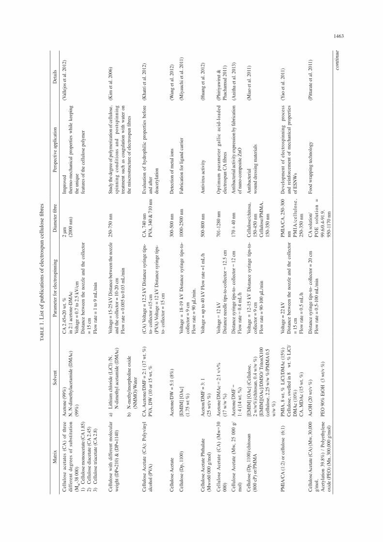
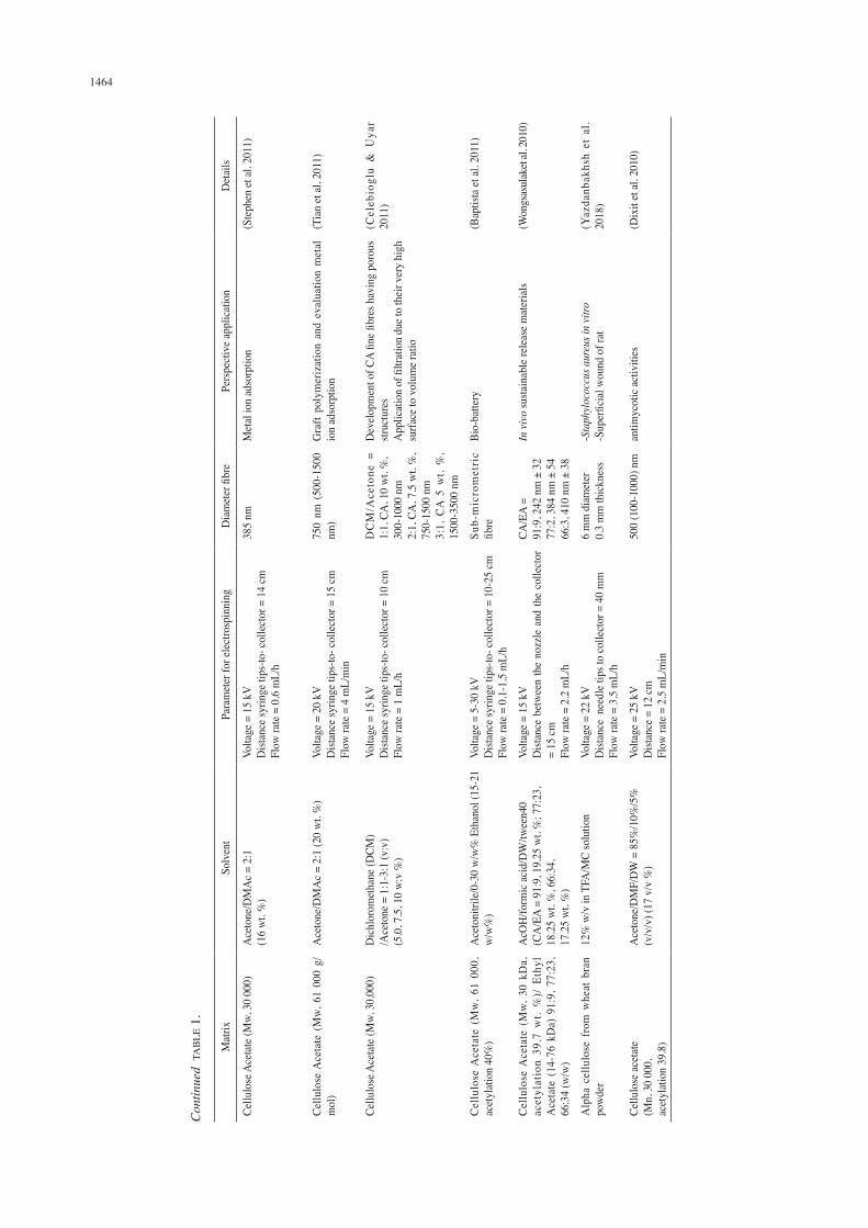
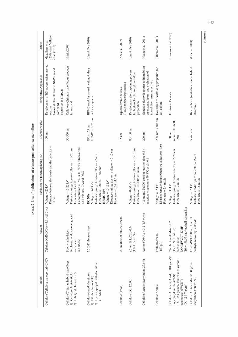
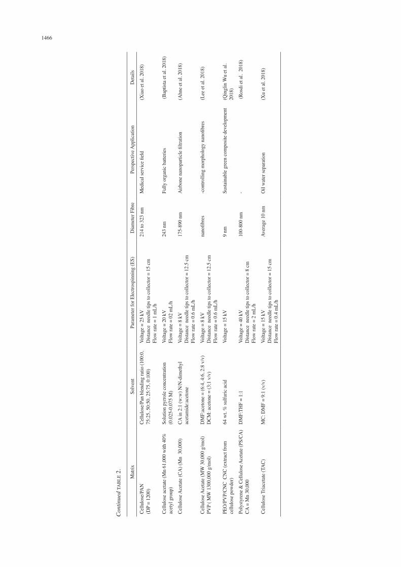
An overall summary list of publications on cellulose fibres and nanofibres mainly from 2006 to 2018 that have been successfully electrospun into super fine fibres is listed in Tables 1 and 2 respectively, with corresponding details including electrospinning solvents, parameters for electrospinning process in different cellulose solution, average diameter of fibres and perspective applications of the corresponding fibres. It was found in the open literature that more than 20 cellulose solutions have been successfully electrospun into fibres with diameters ranging from <30 nm to over 4 mm. Strictly, the terms ‘nanofibres’ and ‘nano-scaled fibres’ are referred to those having average diameters of less than 100 nm (Huang et al. 2003) when at most publications in Table 1 have used the term ‘nanofibres’ for their products even in the cases where the average diameters were 200-1000 nm. Besides, many studies reported in Table 1 included the wording ‘Electrospun cellulose nanofibre’ in their publication headline when in fact the reactant used was cellulose
1463TA
BLE
1. L
ist o
f pub
licat
ions
of e
lect
rosp
un c
ellu
lose
fibr
es
Mat
rixSo
lven
tPa
ram
eter
for e
lect
rosp
inni
ngD
iam
eter
fibr
ePe
rspe
ctiv
e app
licat
ion
Det
ails
Cellu
lose
ace
tate
s (C
A)
of t
hree
di
ffer
ent
degr
ees
of s
ubst
itutio
n (M
W-3
8 00
0)1)
Ce
llulo
se m
onoa
cetat
e (CA
1.85
)2)
Ce
llulo
se d
iace
tate
(CA
2.45
)3)
Ce
llulo
se tr
iace
tate
(CA
2.8)
Ace
tone
(99%
) N
, N-d
imet
hyla
ceta
mid
e (D
MA
c)
(99%
)
CA 2
.45=
20 w
t. %
in 2
:1 ac
eton
e-D
MA
c Vo
ltage
= 0
.7 to
2.5
kV
/cm
D
istan
ce b
etw
een
the
nozz
le a
nd th
e co
llect
or
= 15
cmFl
ow ra
te =
1 to
9 m
L/m
in
2 μm
(200
0 nm
)Im
prov
edth
erm
o-m
echa
nica
l pro
perti
es w
hile
kee
ping
th
e uni
que
feat
ures
of t
he ce
llulo
se p
olym
er
(Val
lejo
s et a
l. 20
12)
Cellu
lose
with
diff
eren
t mol
ecul
ar
wei
ght (
DP=
210)
& (D
P=11
40)
a)
Lith
ium
chlo
ride (
LiCl
) /N
, N
-dim
ethy
l ace
tam
ide (
DM
Ac)
b)
N-m
ethy
lmor
phol
ine o
xide
(N
MM
O)/W
ater
Volta
ge =
15-2
5 kV
Dist
ance
betw
een t
he no
zzle
an
d th
e col
lect
or =
10-
20 cm
Flow
rate
= 0
.005
to 0
.03
mL/
min
250-
750
nmSt
udy t
he de
gree
of po
lym
eriza
tion o
f cell
ulos
e, sp
inni
ng c
ondi
tions
and
po
stsp
inni
ng
treat
men
t suc
h as
coa
gula
tion
with
wat
er o
n th
e misc
rostr
uctu
re o
f ele
ctro
spun
fibr
es
(Kim
et al
. 200
6)
Cellu
lose
Ace
tate
(CA
): Po
lyvi
nyl
alco
hol (
PVA
)CA
, Ace
tone
/DM
F =
2:1
(17
wt.
%)
PVA
, DW
(10
or 1
5 w
t. %
(CA
) Vol
tage
= 1
2.5
kV D
istan
ce sy
ringe
tips
-to
- col
lect
or =
15 cm
(PVA
) Vol
tage
= 1
2 kV
Dist
ance
syrin
ge ti
ps-
to- c
olle
ctor
= 1
5 cm
CA, 7
40 n
mPV
A, 3
40 &
710 n
mEv
alua
tion
of h
ydro
phili
c pr
oper
ties
befo
re
and
afte
r de
acet
ylat
ion
(Kha
tri et
al. 2
012)
Cellu
lose
Ace
tate
Ace
tone
/DW
= 5
:1 (8
%)
300-
500
nmD
etec
tion
of m
etal
ions
(Wan
g et
al. 2
012)
Cellu
lose
(Dp,
110
0)
[EM
IM] [
OA
c]
(1.7
5 w
t %)
Volta
ge =
18-
19 k
V D
istan
ce s
yrin
ge ti
ps-to
- co
llect
or =
9 cm
Flow
rate
= 9
0 μL
/min
.
1000
-250
0 nm
Fabr
icat
ion
for l
igan
d ca
rrier
(Miy
auch
i et a
l. 20
11)
Cellu
lose
Ace
tate
Pht
hala
te
(Mw
=60
000
g/m
ol)
Ace
ton/
DM
F =
3: 1
(2
5 w
t/v %
)Vo
ltage
= u
p to
40
kV F
low
rate
=1
mL/
h50
0-80
0 nm
Ant
iviru
s act
ivity
(Hua
ng et
al. 2
012)
Cel
lulo
se A
ceta
te (
CA
) (M
w=3
0 00
0)A
ceto
ne/D
MA
c = 2
:1 v
/v%
(1
7 w
/w %
)Vo
ltage
= 1
2 kV
D
istan
ce n
eedl
e tip
s-to
-col
lect
or =
12.
5 cm
701-
1280
nm
Opt
imum
par
amet
er g
allic
aci
d-lo
aded
el
ectro
spun
CA
fibre
s (P
hiriy
awiru
t &
Phac
ham
ud 2
011)
Cellu
lose
Ace
tate
(M
w, 2
5 00
0 g/
mol
)A
ceto
ne/D
MF
= 1:
4 (1
4 w
t. %
)D
istan
ce sy
ringe
tips
-to- c
olle
ctor
= 1
2 cm
Flow
rate
= 0
.4 m
L/h
170
± 40
nm
Ant
ibac
teria
l act
ivity
expr
essio
n by f
abric
atio
n of
nan
o-co
mpo
site Z
nO(A
nith
a et a
l. 20
13)
Cellu
lose
(Dp,
110
0)/c
hito
san
(800
cP) o
r/PM
MA
[EM
IM] [
OA
c] (C
ellu
lose
, 2
w/w
%)/c
hito
san,
0.4
w/w
%)
[EM
IM][O
Ac]
/DM
SO/ T
riton
X10
0 (c
ellu
lose
, 2.2
5 w
/w %
/PM
MA
0.5
w/w
%)
Volta
ge =
12-
15 k
V D
istan
ce s
yrin
ge ti
ps-to
- co
llect
or =
9 cm
Flow
rate
= 9
0-10
0 μL
/min
Cellu
lose
/chi
tosa
, 15
0-45
0 nm
Ce
llulo
se/P
MM
A,
150-
350
nm
Ant
ibac
teria
l w
ound
dre
ssin
g m
ater
ials
(Mia
o et
al. 2
011)
PMIA
/CA
(1:2
) or c
ellu
lose
(6:
1)
PMIA
, 8 w
t. %
LiC
l/DM
Ac (
15%
) Ce
llulo
se, s
wel
led
in 8
w
t. %
LiC
l/D
MA
c (10
%)
CA, M
DA
c (15
wt.
%)
Volta
ge =
22
kV
Dist
ance
bet
wee
n th
e no
zzle
and
the
colle
ctor
=
15 cm
Flow
rate
= 0
.5 m
L/h
PMIA
/CA
, 250
-300
nm
PM
IA/c
ellu
lose
, 25
0-35
0 nm
Dev
elop
men
t of
ele
ctro
spin
ning
pr
oces
s an
d re
info
rcem
ent o
f m
echa
nica
l pro
perti
es
of E
SNW
s
(Yao
et al
. 201
1)
Cellu
lose
Ace
tate
(CA
) (M
w, 30
,000
g/
mol
, A
cety
latio
n 39
.8%
) / P
olye
thyl
ene
oxid
e (PE
O) (
Mn,
300
,000
g/m
ol)
AcO
H (2
0 w
t/v %
)
PEO
90%
EtO
H (
3 w
t/v %
)
Dist
ance
syrin
ge ti
ps-to
- col
lect
or =
20
cmFl
ow ra
te =
0.5
-100
mL/
min
CA so
lutio
n/PO
E s
olut
ion
= 99
.6/0
.4-9
1.9,
95
0-11
70 n
m
Food
wra
ppin
g te
chno
logy
(Pitt
arat
e et a
l. 20
11)
cont
inue
1464
Con
tinue
d T
AB
LE 1
.
Mat
rixSo
lven
tPa
ram
eter
for e
lect
rosp
inni
ngD
iam
eter
fibr
ePe
rspe
ctiv
e app
licat
ion
Det
ails
Cellu
lose
Ace
tate
(Mw,
30
000)
Ace
tone
/DM
Ac =
2:1
(1
6 w
t. %
) Vo
ltage
= 1
5 kV
D
istan
ce sy
ringe
tips
-to- c
olle
ctor
= 1
4 cm
Flow
rate
= 0
.6 m
L/h
385
nmM
etal
ion
adso
rptio
n(S
teph
en et
al. 2
011)
Cellu
lose
Ace
tate
(M
w, 6
1 00
0 g/
mol
)A
ceto
ne/D
MA
c = 2
:1 (2
0 w
t. %
) Vo
ltage
= 2
0 kV
D
istan
ce sy
ringe
tips
-to- c
olle
ctor
= 1
5 cm
Flow
rate
= 4
mL/
min
750
nm (
500-
1500
nm
) G
raft
poly
mer
izat
ion
and
eval
uatio
n m
etal
io
n ad
sorp
tion
(Tia
n et
al. 2
011)
Cellu
lose
Ace
tate
(Mw,
30,
000)
D
ichl
orom
etha
ne (D
CM)
/Ace
tone
= 1
:1-3
:1 (v
:v)
(5.0
, 7.5
, 10
w:v
%)
Volta
ge =
15
kV
Dist
ance
syrin
ge ti
ps-to
- col
lect
or =
10
cmFl
ow ra
te =
1 m
L/h
DC
M/A
ceto
ne =
1:
1, C
A, 1
0 w
t. %
, 30
0-10
00 n
m
2:1,
CA
, 7.5
wt.
%,
750-
1500
nm
3:1,
CA
5 w
t. %
, 15
00-3
500
nm
Dev
elop
men
t of C
A fin
e fibr
es h
avin
g po
rous
str
uctu
res
App
licat
ion
of fi
ltrat
ion
due t
o th
eir v
ery
high
su
rface
to v
olum
e rat
io
(Cel
ebio
glu
& U
yar
2011
)
Cel
lulo
se A
ceta
te (
Mw,
61
000,
ac
etyl
atio
n 40
%)
Ace
toni
trile
/0-3
0 w
/w%
Eth
anol
(15-
21
w/w
%)
Volta
ge =
5-3
0 kV
D
istan
ce sy
ringe
tips
-to- c
olle
ctor
= 1
0-25
cmFl
ow ra
te =
0.1
-1.5
mL/
h
Sub-
mic
rom
etri
c fib
reBi
o-ba
ttery
(Bap
tista
et al
. 201
1)
Cel
lulo
se A
ceta
te (
Mw,
30
kDa,
ac
etyl
atio
n 39
.7 w
t. %
)/ E
thyl
A
ceta
te (
14-7
6 kD
a) 9
1:9,
77:
23,
66:3
4 (w
/w)
AcO
H/fo
rmic
acid
/DW
/twee
n40
(CA
/EA
= 91
:9, 1
9.25
wt.
%; 7
7:23
, 18
.25
wt.
%, 6
6:34
, 17
.25
wt.
%)
Volta
ge =
15
kV
Dist
ance
bet
wee
n th
e no
zzle
and
the
colle
ctor
=
15 cm
Flow
rate
= 2
.2 m
L/h
CA/E
A =
91:9
, 242
nm
± 3
2 77
:2, 3
84 n
m ±
54
66:3
, 410
nm
± 3
8
In vi
vo su
stain
able
rele
ase m
ater
ials
(Won
gsas
ulak
et al.
2010
)
Alp
ha c
ellu
lose
fro
m w
heat
bra
n po
wde
r12
% w
/v in
TFA
/MC
solu
tion
Volta
ge =
22
kV
Dist
ance
nee
dle t
ips t
o co
llect
or =
40
mm
Flow
rate
= 3
.5 m
L/h
6 m
m d
iam
eter
0.3
mm
thic
knes
s-S
taph
yloc
occu
s aur
eus i
n vi
tro-S
uper
ficia
l wou
nd o
f rat
(Yaz
danb
akhs
h et
al.
2018
)
Cellu
lose
acet
ate
(Mn,
30
000,
ac
etyl
atio
n 39
.8)
Ace
tone
/DM
F/D
W =
85%
/10%
/5%
(v
/v/v
) (17
v/v
%)
Volta
ge =
25
kV
Dist
ance
= 1
2 cm
Flow
rate
= 2
.5 m
L/m
in
500
(100
-100
0) n
man
timyc
otic
activ
ities
(Dix
it et
al. 2
010)
1465
TAB
LE 2
. Lis
t of p
ublic
atio
ns o
f ele
ctro
spun
cel
lulo
se n
anofi
bres
Mat
rixSo
lven
tPa
ram
eter
for E
lect
rosp
inni
ng (E
S)D
iam
eter
Fib
rePe
rspe
ctiv
e App
licat
ion
Det
ails
Cellu
lose
/Cel
lulo
se n
anoc
rysta
l (CN
C)Ce
llulo
se, N
MM
O/DW
= 4:
1 wt (
1.5 w
t )Vo
ltage
= 2
0 kV
D
istan
ce b
etw
een
the n
ozzl
e and
the c
olle
ctor
=
10 cm
100
nmD
evel
opm
ent o
f ES
proc
ess u
sing
laye
red
nozz
les
havi
ng sh
ell (
cellu
lose
in N
MM
O) a
nd
core
(CN
C di
sper
sion
in D
MSO
)
Mag
alha
es et
al.
(200
9) an
d V
alle
jos
et al
. (20
12)
Cellu
lose
/Chi
tosa
n hy
brid
nan
ofibr
es1)
Ce
llulo
se A
ceta
te (C
A)
2)
Dib
utyr
yl ch
itin
(DBC
)
Buty
ric an
hydr
ide,
Perc
hlor
ic ac
id, a
ceto
ne, g
laci
al
acet
ic ac
idan
d D
MA
c
Volta
ge =
15-
25 k
V
Dist
ance
syrin
ge ti
ps-to
- col
lect
or =
15-
20 cm
Flow
rate
= 1
mL/
hCo
ncen
tratio
n =
15%
in 1
/1 w
/w ac
eton
e/ac
etic
ac
id m
ixtu
re, C
A an
d D
BC
30-3
50 n
mCe
llulo
se-C
hito
san
nano
fibro
us p
rodu
cts
for m
edic
alH
sieh
(200
9)
Cellu
lose
-bas
ed N
anofi
bres
1)
Ethy
l-cel
lulo
se (E
C)
2)
Hyd
roxy
prop
yl m
ethy
lcel
lulo
se
(HPM
C)
2,2,
2-Tr
ifluo
roet
hano
lEC
NFs
Volta
ge =
5-2
8 kV
Dist
ance
syrin
ge ti
ps-to
- col
lect
or =
5 cm
Flow
rate
= 0
.001
-0.0
1 m
L/m
inH
PMC
NFs
Volta
ge =
10-
15 k
V
Dist
ance
syrin
ge ti
ps-to
- co
llect
or =
5-1
5 cm
Flow
rate
= 0
.05
mL/
min
EC =
155
nm
HPM
C =
162
nmH
PMC
used
for w
ound
hea
ling
& d
rug
deliv
ery
syste
m(L
im &
Pyo
201
0)
Cellu
lose
(woo
d)2:
1 m
ixtu
re o
f tol
uene
/eth
anol
15 n
mO
ptoe
lect
roni
c dev
ices
,Ti
ssue
engi
neer
ing
scaf
fold
(Abe
et al
. 200
7)
Cellu
lose
(Dp,
120
00)
8.5
wt.
% L
iCl/D
MA
c (1
.0-1
.35
wt.
%)
Volta
ge =
0-3
0 kV
D
istan
ce sy
ringe
tips
-to- c
olle
ctor
= 1
0-15
cmFl
ow ra
te =
0.0
1-0.
06 m
L/m
in
80-1
00 n
mD
evel
opm
ent e
lect
rosp
inni
ng p
roce
ss
for h
igh
mol
ecul
ar w
eigh
t cel
lulo
se
prep
arat
ion
(Lim
& P
yo 2
010)
Cellu
lose
Ace
tate
(ace
tyla
tion,
29.
6%)
Ace
tone
/DM
Ac =
3:2
(15
wt %
) 4.
2 m
g/m
L N
aIO
4 co
nten
t rea
ctio
n tim
e 6.8
h
reac
tion
tem
pera
ture
30.
8°C
at p
H 6
.120
0 nm
Gen
erat
e ald
ehyd
e gro
ups t
o im
mob
ilize
an
enzy
me,
lipas
e, an
d ev
alua
tion
of
imm
obili
zed
enzy
me a
ctiv
ity
(Hua
ng et
al. 2
011)
Cellu
lose
Ace
tate
Tr
ifluo
roet
hano
l(1
50 g
/L)
Volta
ge =
15
kV
Dista
nce b
etwee
n the
nozz
le an
d the
colle
ctor =
10 cm
Flow
rate
= 2
.4 m
L/h
200
nm-3
000
nm
Eval
uatio
n of
scaf
fold
ing
prop
ertie
s for
ce
ll cu
lture
(Fili
on et
al.
2011
)
Cellu
lose
Ace
tate
(CA
) (d,
1.1
84 g
/cm
3 )/ Po
ly (a
cryl
onitr
ile) (
PAN
) (D
, 1.1
84 g
/cm
3 ) / m
ultiw
alle
d ca
rbon
na
notu
bes (
MW
CNT)
(D
, 1.2
-1.7
g/c
m3 )
CA, A
ceto
ne/D
MA
c =1:
2 (1
5 w
t. %
/10
wt.
%),
core
solu
tion
PAN
/MW
CNT,
DM
F (1
0 w
t. %
/35
wt.
%),
shel
l sus
pens
ion
Volta
ge =
30
kV
Dist
ance
syrin
ge ti
ps-to
-col
lect
or =
15-
20 cm
Flow
rate
= 0
.2 m
L/h
150
nm(c
ore,
~60;
shel
l, ~5
0)
Elec
troni
c Dev
ices
(Lisu
nova
et al
. 201
0)
Cellu
lose
Ace
tate
(Mw
30,
000g
/mol
, ac
etyl
atio
n 39
.8 w
t. %
)1:
1=D
MSO
:TH
F +
0.1
wt.
%
diet
hyla
min
o et
hyl c
hlor
ide
Volta
ge =
15
kV
Dist
ance
nee
dle t
ips t
o co
llect
or =
25
cmFl
ow ra
te =
0.8
mL/
h
58-6
0 nm
Bio-
synt
hesis
(mut
i-dim
ensio
nal h
ybrid
str
uctu
re)
(Lv
et al
. 201
8) cont
inue
1466
Con
tinue
d TA
BLE
2.
Mat
rixSo
lven
tPa
ram
eter
for E
lect
rosp
inni
ng (E
S)D
iam
eter
Fib
rePe
rspe
ctiv
e App
licat
ion
Det
ails
Cellu
lose
/PA
N
(D
P =
1200
)Ce
llulo
se/P
an b
lend
ing
ratio
(100
:0,
75:2
5, 5
0:50
, 25:
75, 0
:100
)Vo
ltage
= 2
5 kV
D
istan
ce n
eedl
e tip
s to
colle
ctor
= 1
5 cm
Flow
rate
= 1
mL/
h
214
to 3
23 n
mM
edic
al se
rvic
e fiel
d(X
iao
et al
. 201
8)
Cellu
lose
acet
ate (
Mn
61,0
00 w
ith 4
0%
acet
yl g
roup
)So
lutio
n py
rrole
conc
entra
tion
(0
.025
-0.0
75 M
)Vo
ltage
= 2
0 kV
Fl
ow ra
te =
02
mL/
h24
3 nm
Fully
org
anic
bat
terie
s (B
aptis
ta et
al. 2
018)
Cellu
lose
Ace
tate
(CA
) (M
n 3
0,00
0)CA
in 2
:1 (w
:w) N
/N-d
imet
hyl
acet
amid
e:ac
eton
eVo
ltage
= 8
kV
D
istan
ce n
eedl
e tip
s to
colle
ctor
= 1
2.5
cmFl
ow ra
te =
0.6
mL/
h
175-
890
nmA
irbon
e nan
opar
ticle
filtr
atio
n(A
hne e
t al.
2018
)
Cellu
lose
Ace
tate
(MW
30,
000
g/m
ol)
PVP
( MW
130
0,00
0 g/
mol
)D
MF:
acet
one =
(6:4
, 4:6
, 2:8
v/v
)D
CM: a
ceto
ne =
(3;1
v/v
)Vo
ltage
= 8
kV
D
istan
ce n
eedl
e tip
s to
colle
ctor
= 1
2.5
cmFl
ow ra
te =
0.6
mL/
h
nano
fibre
s-c
ontro
lling
mor
phol
ogy
nano
fibre
s(L
ee et
al. 2
018)
PEO
/PV
P/CN
C C
NC
(ext
ract
from
ce
llulo
se p
owde
r)64
wt.
% su
lfuric
acid
Volta
ge =
15
kV
9 nm
Susta
inab
le g
reen
com
posit
e dev
elop
men
t(Q
ingl
in W
u et
al.
2018
)
Poly
styre
ne &
Cel
lulo
se A
ceta
te (P
S/CA
)CA
= M
n 30
,000
DM
F:TH
F =
1:1
Volta
ge =
40
kV
Dist
ance
nee
dle t
ips t
o co
llect
or =
8 cm
Flow
rate
= 2
mL/
h
100-
800
nm-
(Ros
di et
al.
2018
)
Cellu
lose
Tria
ceta
te (T
AC)
MC:
DM
F =
9:1
(v/v
)Vo
ltage
= 1
5 kV
D
istan
ce n
eedl
e tip
s to
colle
ctor
= 1
5 cm
Flow
rate
= 0
.4 m
L/h
Aver
age 1
0 nm
Oil
wat
er se
para
tion
(Xu
et al
. 201
8)
1467
diameter ranging. In addition, using chemomechanical isolation, Wang and Sain (2007) synthesised soybean stock-based nanofibres with a diameter ranging from 50-100 nm. Then, X-ray crystallography was accomplished to investigate the percentage crystallinity after various stages of chemomechanical treatment. It has been found that samples crystallinity increased after each stage of nanofiber development. The nanofibre suspension achieved after the high pressure defibrillation was analysed to determine diameters using AFM. The AFM image displays the surface of air dried soybean stock nanofibres. It was seen that the fibres were nanosized with diameter of nanofibres within the range of 50-100 nm.
NANOCELLULOSE APPLICATION
Biomedical Application Silver nanoparticles combined with cellulose nanofibers maybe connected as antibacterial specialists in wound dressing, swathes, inserts, medicaments as well as in antimicrobial nourishment pressing, face veils, water cleaning frameworks and air channel products (Kozlowski et al. 2007; Laborie & Brown 2009). The discoverer expressed that nanocomposite included by silver nanoparticle and cellulose nanofibre considered natural cordial and the planning technique is straightforward and flexible for scaling (Chandrabhas 2008; Moon et al. 2011). The blend of nanocellulose and silver nanoparticles can be utilised for microbicidal medicaments. Cellulose spinning solution mixed with silver nanoparticles can serve as bacteriostatic, bactericidal and fungicidal. Cellulose nanofibres prepared with nanosilver have been observed to be productive across Staphylococcus aureous amid the bacterial development process (Moon et al. 2011). Essentially, oxidised carboxyl groups in cellulose nanofibres have many applications for antibacterial activity. Along these lines, E. coli as the representative exhibited microbicidal activity for water purification procedure in view of an oxidised cellulose nanofibrous film (Benjamin et al. 2010). In 2009, Anderson depicted the employment of nanocellulose for skin treatment in the conveyance of makeup and medicaments (Andersen 2009). The mix with the first layer comprises biomaterial including cellulose while the second layer is made out of polymer nanofibres that contain the dynamic cosmetic content similar to a vitamin or peptide (Andersen 2009). Additionally, nanocellulose can be applied for cosmetic treatments including the evacuation of cosmetics, cleaning of skin and hair as well as the treatment for dry or delicate skin using cellulose film made up from oil in water emulsion without surfactants. According to Rebecca Davies, it has been reported that cellulose nanoparticle hydrogels and aerogels with normal pore distances across of under 100 nm can be connected in a few biomedical applications such as biosensors, platforms for tissue engineering, pathogen recognition, drug conveyance process, contact focal points, medical electrodes, breast embed, therapeutic gadgets and biocatalysts (Thielemans & David 2011). Anna et al. (2007)
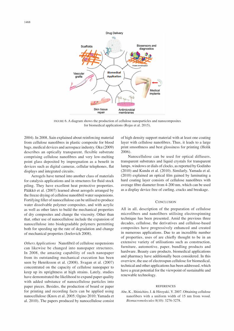
have contemplated various novel materials considering cellulose nanofibres such as froths, nanopapers, aerogels and composite of starch nanocellulose. Henriksson et al. (2007) produced cellulose nanofibres from wood mash and amylopectin rich potato starch for biofoams application. Many investigators are interested in applications of biomedical and packaging material using the concept of cellulose nanocomposite biofoams. Nanocellulose in its naturally feature state is unharmed to people and biocompatible. Therefore, nanocellulose can be utilised for health care use like personal hygiene products, biomedicines and cosmetics. Besides, nanocellulose dispersions can be accounted for stabilisation of medical suspensions against phase separation and sedimentation of huge contents. From a view of chemical, immobilisation of enzymes and other drugs can be utilised by mutated cellulose as a promising carrier. For example, bearer drug complex can permeate through skin pores and treat skin infections due to its nano size molecule and can be connected as a peeling operator in beautifying agent products (Ponomarenko et al. 2008). Figure 6 shows the production of cellulose nanoparticles and nanocomposites for biomedical applications (Rojas et al. 2015).
Technical Applications Cellulose has a unique feature and can be transformed in many different forms such as crystallisation, nanocrystals, whiskers, nanofibrils, and nanofibres that are usually used in many technical applications. Nanocellulose combines fascinating physical and morphological properties such as large surface area, hydrophilicity, sites for chemical modifications, formation of semicrystalline fibres that allow numerous applications (John & Thomas 2008). Normally, filtration membranes can be applied using cellulose nanofibres (Benjamin et al. 2010, 2008; Koslow 2003). According to Benjamin (2010), the electrospinning technology for preparing cellulose nanofibrous membranes with average fibre diameter of 100 nm, homogeneous handling and the ability for surface functionalisation has significantly improved water flow and can be used as microfiltration membranes. These are beneficial for low pressure-microfiltration, nano filtration, ultra-filtration and forward osmosis. Moreover, Koslow (2003) expressed that cellulose nanofibre filter contains a microbiological interception enhancing agent consisting biologically active metal with the ability of imparting a positive charge at the very least portion of micropores. Cellulose surface chemical modifications are the encouraging strategies to enhance mechanical and thermal properties of varied polymers such as elongation at break, tensile strength at break, biodegradability and water-retention for technical applications (John & Thomas 2008; Vallejos et al. 2012). Correspondingly, cellulose nanofibres surface modified can be used in coating, filters, membranes, adhesives, sealings, packing and cosmetics (Bordeanu & Eyholzer 2009). Other technical applications for nanocellulose emerged up on its existence in composites for reinforced biomaterials (Oksman et al. 2006; Sain
1468
2004). In 2008, Sain explained about reinforcing material from cellulose nanofibres in plastic composite for blood bags, medical devices and aerospace industry. Oku (2009) describes an optically transparent, flexible substrate comprising cellulose nanofibres and very low-melting point glass deposited by impregnation as a benefit in devices such as digital cameras, cellular telephones, flat displays and integrated circuits. Aerogels have turned into another class of materials for catalysis applications and in structures for fluid stock piling. They have excellent heat protective properties. Paakkö et al. (2007) learned about aerogels arranged by the freeze drying of cellulose nanofibril water suspensions. Fortifying filler of nanocellulose can be utilised to produce water dissolvable polymer composites, and with acrylic as well as other latex to build the mechanical properties of dry composites and change the viscosity. Other than that, other use of nanocellulose include the expansion of nanocellulose into biodegradable polymers permitting both for speeding up the rate of degradation and change of mechanical properties (Ioelovich 2008).
Others Applications Nanofibril of cellulose suspensions can likewise be changed into nanopaper structures. In 2008, the amazing capability of such nanopaper from its outstanding mechanical execution has been seen by Henriksson et al. (2008). Svagan et al. (2007) concentrated on the capacity of cellulose nanopaper to keep up its uprightness at high strains. Lately, studies have demonstrated the likelihood to expand paper quality with added substance of nanocellulose particles into paper pieces. Besides, the production of board or paper for printing and recording facts can be applied using nanocellulose (Knox et al. 2005; Ogino 2010; Yamada et al. 2010). The papers produced by nanocellulose consist
of high density support material with at least one coating layer with cellulose nanofibres. Thus, it leads to a large print smoothness and best glossiness for printing (Holik 2006). Nanocellulose can be used for optical diffusers, transparent substrates and liquid crystals for transparent lamps, windows or dials of clocks, as reported by Godinho (2010) and Kundu et al. (2010). Similarly, Yamada et al. (2010) explained an optical film gained by laminating a hard coating layer consists of cellulose nanofibres with average fibre diameter from 4-200 nm, which can be used as a display device free of curling, cracks and breakage.
CONCLUSION
All in all, description of the preparation of cellulose microfibers and nanofibers utilising electrospinning technique has been presented. Amid the previous three decades, cellulose, the derivatives and cellulose-based composites have progressively enhanced and created in numerous applications. Due to an incredible number of properties, uses of are chiefly thought to be in an extensive variety of utilisations such as construction, furniture, automotive, paper, bundling products and hardware. Beauty care products, biomedical applications and pharmacy have additionally been considered. In this overview, the use of electrospun cellulose for biomedical, technical and other applications has been addressed, which have a great potential for the viewpoint of sustainable and renewable technology.
REFERENCES
Abe, K., Shinichiro, I. & Hiroyuki, Y. 2007. Obtaining cellulose nanofibers with a uniform width of 15 nm from wood. Biomacromolecules 8(10): 3276-3278.
FIGURE 6. A diagram shows the production of cellulose nanoparticles and nanocomposites for biomedical applications (Rojas et al. 2015).
1469
Ahne, J., Qinghai, L., Eric, C. & Zhongchao, T. 2018. Electrospun cellulose acetate nanofibers for airborne nanoparticle filtration. Textile Research Journal. https://doi.org/10.1177/0040517518807440.
Akihito, O. 2010. Papier pour enregistrement d’informations et papier traité. doi: WO 2011001706 A1.
Alemdar, A. & Sain, M. 2008a. Biocomposites from wheat straw nanofibers: Morphology, thermal and mechanical properties. Composites Science and Technology 68(2): 557-565. doi:10.1016/j.compscitech.2007.05.044.
Alemdar, A. & Sain, M. 2008b. Isolation and characterization of nanofibers from agricultural residues - Wheat straw and soy hulls. Bioresource Technology 99(6): 1664-1671. doi:10.1016/j.biortech.2007.04.029.
Anitha, S., Brabu, B., John, D., Thiruvadigal, Gopalakrishnan, C. & Natarajan, T.S. 2013. Optical, bactericidal and water repellent properties of electrospun nano-composite membranes of cellulose acetate and ZnO. Carbohydrate Polymers 97(2): 856-863. https://doi.org/10.1016/j.carbpol.2013.05.003.
Anderson, E. 2009. Skin care compositions for the delivery of agents. WO2010115426 A1.
Anna, J.S., My, A.S.A.S. & Berglund, L.A. 2007. Biomimetic polysaccharide nanocomposites of high cellulose content and high toughness. Biomacromolecules 8(8): 2556-2563. doi:10.1021/BM0703160.
Baptista, A.C., Ropio, I., Romba, B., Nobre, J.P., Henriques, C., Silva, J.C., Martins, J.I., Borges, J.P. & Ferreira, I. 2018. Cellulose-based electrospun fibers functionalized with polypyrrole and polyaniline for fully organic batteries. Journal of Materials Chemistry. 6(1): 256-265. https://doi.org/10.1039/c7ta06457h.
Baptista, A.C., Martins, J.J., Fortunato, E., Martins, R., Borges, J.P. & Ferreira, I. 2011. Thin and flexible bio-batteries made of electrospun cellulose-based membranes. Biosensors and Bioelectronics 26(5): 2742-2745. https://doi.org/10.1016/j.bios.2010.09.055.
Benjamin, C., Benjamin, H. & Hongyang, M. 2010. High flux high efficiency nanofiber membranes and methods of production thereof. USWO2010042647A2.
Benjamin, C., Benjamin H. & Hongyang, M. 2008. Membranes de séparation de fluides à haut flux comprenant une couche de cellulose ou d’un dérivé de cellulose. WO2009025900 A2.
Bledzki, A.K., Reihmane, S. & Gassan, J. 1996. Properties and modification methods for vegetable fibers for natural fiber composites. Journal of Applied Polymer Science 59(8): 1329-1336. doi:10.1002/(SICI)1097-4628(19960222)59:8<1329::AID-APP17>3.3.CO;2-5.
Bognitzki, M., Czado, W., Frese, T., Schaper, A., Hellwig, M., Steinhart, M., Greiner, A. & Wendorff, J.H. 2001. Nanostructured fibers via electrospinning. Advanced Materials 13(1): 70-72. doi:10.1002/1521-4095(200101)13:1<70::AID-ADMA70>3.0.CO;2-H.
Bordeanu, N., Eyholzer, C. & Zimmermann, T. 2009. Surface modified cellulose nanofibers. doi:WO 2010066905 A1.
Celebioglu, A. & Uyar, T. 2011. Electrospun porous cellulose acetate fibers from volatile solvent mixture. Materials Letters 65(14): 2291-2294. https://doi.org/10.1016/j.matlet.2011.04.039.
Chandrabhas, N. 2008. Nanoparticle composition and process thereof. US8834917B2.
Cucolo, J.A., Aminuddin, N. & Frey, M. 2001. Structure formation in polymeric fibers. Hanser Gardner Publications
http://www.hanserpublications.com/Products/227-structure-formation-in-polymeric-fibers.aspx. pp. 296-328.
de Morais Teixeira, E., Corrêa, A.C., Manzoli, A., de Lima Leite, F., de Oliveira, C.R. & Mattoso, L.H.C. 2010. Cellulose nanofibers from white and naturally colored cotton fibers. Cellulose 17(3): 595-606. doi:10.1007/s10570-010-9403-0.
Dixit, V., Jagdish, T. & Kay, O.S. 2010. Fungal growth inhibition of regenerated cellulose nanofibrous membranes containing quillaja saponin. Archives of Environmental Contamination and Toxicology 59(3): 417-423. https://doi.org/10.1007/s00244-010-9493-6.
Faten Ermala Che Othman, Norhaniza Yusof, Amirul Afiat Raffi, Hasrinah Hasbullah, Farhana Aziz, Wan Norharyati Wan Salleh & Ahmad Fauzi Ismail. 2017. Preparation and characterization of different loading of zinc oxide on activated carbon nanofibers. Malaysian Journal of Analytical Science 21(2): 365-371. doi:10.17576/mjas-2017-2102-11.
Fauzee, S.N. & Othaman, R. 2013. Extraction and dissolution of cellulose from nypa fruit husk for nanofibers fabrication. AIP Conference Proceedings 1571 (December 2013): 904-910. doi:10.1063/1.4858769.
Filion, T.M., Artem, K. & Jie, S. 2011. Chemically modified cellulose fibrous meshes for use as tissue engineering scaffolds. Bioorganic and Medicinal Chemistry Letters 21(17): 5067-5070. https://doi.org/10.1016/j.bmcl.2011.04.032.
Freire, M.G., Ana, R., Teles, R., Rute, A.S., Ferreira, L.D., Carlos, J.A., Lopes-da-Silva & João, A.P.C. 2011. Electrospun nanosized cellulose fibers using ionic liquids at room temperature. Green Chemistry 13(11): 3173. https://doi.org/10.1039/c1gc15930e.
Frey, M. & Joo, Y. 2004. Cellulose solution in novel solvent and electrospinning thereof. doi:US 20050247236 A1.
Georgopoulos, S.T., Tarantili, P.A., Avgerinos, E., Andreopoulos, A.G. & Koukios, E.G. 2005. Thermoplastic polymers reinforced with fibrous agricultural residues. Polymer Degradation and Stability 90(2 SPEC. ISS.): 303-312. doi:10.1016/j.polymdegradstab.2005.02.020.
Henriksson, M., Berglund, L.A., Isaksson, P., Lindström, T. & Nishino, T. 2008. Cellulose nanopaper structures of high toughness. Biomacromolecules 9(6): 1579-1585. doi:10.1021/bm800038n.
Henriksson, M., Henriksson, G., Berglund, L.A. & Lindström, T. 2007. An environmentally friendly method for enzyme-assisted preparation of microfibrillated cellulose (MFC) nanofibers. European Polymer Journal 43(8): 3434-3441. doi:10.1016/j.eurpolymj.2007.05.038.
Holik, H. 2006. Handbook of Paper and Board. New York: Wiley-VCH.
Hornsby, P.R., Hinrichsen, E. & Tarverdi, K. 1997. Preparation and properties of polypropylene composites reinforced with wheat and flax straw fibres Part II analysis of composite microstructure and mechanical properties JMS60060 JMS60060. Journal of Materials Science 32: 1009-1015. doi:10.1023/A:1018578322498.
Hsieh, J.D.Y. 2009. Cellulose/Chitosan hybrid nanofibers from electrospinning of their ester derivatives. Cellulose 16(2): 247-260. https://doi.org/10.1007/s10570-008-9266-9.
Huang, C., Stefaan, J.S., Ellen, V.G., Guido, V., Joanna, R., Serge, V.C., Chris, V., Coenye, T., Verstraelen, H., Temmerman, M., Demeester, J. & De Smedt, S.C. 2012. Biomaterials electrospun cellulose acetate phthalate fibers for semen induced anti-HIV vaginal drug delivery. Biomaterials 33(3): 962-969. https://doi.org/10.1016/j.biomaterials.2011.10.004.
1470
Huang, X.J., Peng, C.C., Fu, H., Yang, O., Ming, R.C. & Zhi, K.X. 2011. Immobilization of Candida rugosa lipase on electrospun cellulose nanofiber membrane. Journal of Molecular Catalysis B: Enzymatic 70(3-4): 95-100. https://doi.org/10.1016/j.molcatb.2011.02.010.
Huang, Z.M., Zhang, Y.Z., Kotaki, M. & Ramakrishna, S. 2003. A review on polymer nanofibers by electrospinning and their applications in nanocomposites. Composite Science and Technology 63(5): 2223-2253. doi:10.1016/S0266-3538(03)00178-7.
Hutmacher, D.W. & Dalton, P.D. 2011. Melt electrospinning. Chemistry - An Asian Journal 6(1): 44-56. doi:10.1002/asia.201000436.
Ioelovich, M. 2008. Cellulose as a nanostructured polymer. BioResources 3(4): 1403-1418.
Javadian, M., Rostamizadeh, K. & Danafar, H. 2012. Preparation and characterization of electrospinning PEG-PLA nanofibers for sustained release of tamoxifen. Research in Pharmaceutical Sciences 7: 5.
Jaworek, A., Krupa, A., Lackowski, M., Sobczyk, A.T., Czech, T., Ramakrishna, S., Sundarrajan, S. & Pliszka, D. 2009. Electrospinning and electrospraying techniques for nanocomposite non-woven fabric production. Fibres and Textiles in Eastern Europe 75(4): 77-81.
John, M.J. & Thomas, S. 2008. Biofibres and biocomposites. Carbohydrate Polymers 71(3): 343-364. doi:10.1016/j.carbpol.2007.05.040.
Kalia, S., Kaith, B.S. & Vashistha, S. 2011. Cellulose nanofibers reinforced bioplastics and their applications. Handbook of Bioplastics and Biocomposites Engineering Applications, edited by Pilla, S. Hoboken, New Jersey: John Wiley & Sons, Inc. pp. 452-470. doi:10.1002/9781118203699.ch16.
Kaushik, A., Singh, M. & Verma, G. 2010. Green nanocomposites based on thermoplastic starch and steam exploded cellulose nanofibrils from wheat straw. Carbohydrate Polymers 82(2): 337-345. doi:10.1016/j.carbpol.2010.04.063.
Khatri, Z., Kai, W., Byoung-suhk, K. & Ick-soo, K. 2012. Effect of deacetylation on wicking behavior of co-electrospun cellulose acetate/polyvinyl alcohol nanofibers blend. Carbohydrate Polymers 87(3): 2183-2188. https://doi.org/10.1016/j.carbpol.2011.10.046.
Kim, C., Dae-sik, K., Seung-yeon, K., Manuel, M. & Yong, L. 2006. Structural studies of electrospun cellulose nanofibers. Polymer 47(14): 5097-5107. https://doi.org/10.1016/j.polymer.2006.05.033.
Klemm, D., Heublein, B., Fink, H.P. & Bohn, A. 2005. Cellulose: Fascinating biopolymer and sustainable raw material. Angewandte Chemie (International ed. in English) 44(22): 3358-3393. doi:10.1002/anie.200460587.
Knox, D., Klein, E., Babinsky, V. & Klein, S. 2005. Process and apparatus for coating paper. US20070148365A1.
Koslow, E. 2003. Microporous filter media, filtration systems containing same, and methods of making and using. doi:US 20030205530 A1.
Kozlowski, R., Laszkiewicz, B., Kulpinski, P., Muzyczek, M., Czarnecki, P., Rubacha, M., Niekraszewicz, B., Jedrzejczak, J. & Peczek, B. 2007. Method of manufacturing silver nanoparticles, cellulosic fibers and nanofibers containing silver nanoparticles, fibers and nanofibers containing silver nanoparticles, use of silver nanoparticles to the manufacture of cellulosic fibers and nanofibers. European Patent EP0905289.
Krassig, H.A. 1993. Cellulose: Structure, Accessibility, and Reactivity. Philadephia: Gordon and Breach Publishers.
Kundu, S., Feio, G., Pinto, L.F.V., Almeida, P.L., Figueirinhas, J.L. & Godinho, M.H. 2010. Deuterium NMR study of orientational order in cellulosic network microfibers. Macromolecules 43(13): 5749-5755. doi:10.1021/ma100882w.
Laborie, M.P. & Brown, E. 2009. Method of in situ bioproduction and composition of bacterial cellulose nanocomposites. doi:US20090192264 A1.
Leach, M.K., Feng, Z.Q., Tuck, S.J. & Corey, J.M. 2011. Electrospinning fundamentals: optimizing solution and apparatus parameters. Journal of Visualized Experiments (47): e2494-e2494. doi:10.3791/2494.
Lee, H., Masayoshi, N., Daewon, S. & Jung, S. 2018. Control of the morphology of cellulose acetate nanofibers via electrospinning. Cellulose. https://doi.org/10.1007/s10570-018-1744-0.
Lim, Y. & Pyo, J. 2010. Preparation of cellulose-based nanofibers using electrospinning. Intech Open. February. https://www.intechopen.com/books/nanofibers/preparation-of-cellulose-based-nanofibers-using-electrospinning.
Lisboa, U.N.D.E. 2009. Nanofibras celulósicas obtidas por electrospinning a partir 24(11).
Lisunova, M., Attila, H., Benjamin, H., Vitaliy, D. & Stephanie, R. 2010. Nanofibres of CA/PAN with high amount of carbon nanotubes by core-shell electrospinning. Composites Science and Technology 70(11): 1584-1588. https://doi.org/10.1016/j.compscitech.2010.07.001.
Lv, P., Muhammad, N., Qufu, W., Huimin, Z. & Tayyab, N. 2018. A novel in situ self-assembling fabrication method for bacterial cellulose-electrospun nanofiber hybrid structures. Polymers 10(7): 712. https://doi.org/10.3390/polym10070712.
Magalhaes, W.L.E., Cao, X. & Lucia, A.L. 2009. Cellulose nanocrystals/Cellulose core-in-shell nanocomposite assemblies. Langmuir 25(22): 13250-13257. https://doi.org/10.1021/la901928j.
Marsh, J.T. & Wood, F.C. 1942. An Introduction to the Chemistry of Cellulose. London: Chapman & Hall. http://www.archive.org/details/introductiontoth029129mbp.
Medronho, B. & Lindman, B. 2015. Brief overview on cellulose dissolution/regeneration interactions and mechanisms. Advances in Colloid and Interface Science 222: 502-508. doi:10.1016/j.cis.2014.05.004.
Medronho, B., Romano, A., Miguel, M.G., Stigsson, L. & Lindman, B. 2012. Rationalizing cellulose (in)solubility: Reviewing basic physicochemical aspects and role of hydrophobic interactions. Cellulose 19(3): 581-587. doi:10.1007/s10570-011-9644-6.
Meier, H. 1962. Chemical and morphological aspects of the fine structure of wood. Pure App. Chem. 5: 37-52.
Miao, J., Ravindra, C., Pangule, E.E., Paskaleva, Elizabeth, E., Hwang, Ravi, S., Kane, R.J., Linhardt & Jonathan, S.D. 2011. Lysostaphin-functionalized cellulose fibers with antistaphylococcal activity for wound healing applications. Biomaterials 32(36): 9557-9567. https://doi.org/10.1016/j.biomaterials.2011.08.080.
Miyauchi, M., Jianjun, M., Trevor, J.S., Jonathan, S.D. & Robert, J.L. 2011. Chromatography flexible electrospun cellulose fibers as an affinity packing material for the separation of bovine serum albumin. Journal of Chromatography &
1471
Separation Techniques 2: 110. https://doi.org/10.4172/2157-7064.1000110.
M. Sain, M. & Bhatnagar, A. 2004. Manufacturing process of cellulose nanofibers from renewable feed stocks. US 20080146701 A1.
Moon, R.J., Martini, A., Nairn, J., Simonsen, J. & Youngblood, J. 2011. Cellulose nanomaterials review: Structure, properties and nanocomposites. Chem. Soc. Rev. 40: 3941-3994. doi:10.1039/c0cs00108b.
Muhammad Hariz Othman, Mahathir Mohamed, Ibrahim Abdullah & Dahlan Haji Mohd. 2014. Preparation of non-woven fiber mats by mixture of pvc and epoxidized natural rubber. Journal of Nuclear and Related Technology 11(1): 1-10. http://www.nuklearmalaysia.org/publication/jnrt/8-jnrt/9-jnrt-volume-14-2020.
Muhammad Johan Iskandar Zahari, Noraisah Mohd Jahi, Nurul Hanisah Mohd, Ishak Ahmad, Azizah Baharum, Azwan Mat Lazim, Suria Ramli & Rizafizah Othaman. 2018. Enhanced performance of cellulose from palm oil empty fruit bunch (EFB) via acetylation and silylation. Preprints (July): 20. doi:10.20944/preprints201807.0314.v1.
Ohkawa, K. 2015. Nanofibers of cellulose and its derivatives fabricated using direct electrospinning. Molecules 20(5): 9139-9154. doi:10.3390/molecules20059139.
Oksman, K., Bondeson, D. & Syre, P. 2006. Nanocomposites based on cellulose whiskers and cellulose plastics. US20080108772A1.
Oku, Y. 2009. Flexible substrate and manufacturing method thereof. doi:US 20090202843 A1.
Paakkö, M., Ankerfors, M., Kosonen, H., Nykanen, A., Ahola, S., Österberg, M., Ruokolainen, J., Laine, J., Larsson, P.T., Ikkala, O. & Lindström, T. 2007. Enzymatic hydrolysis combined with mechanical shearing and high-pressure homogenization for nanoscale cellulose fibrils and strong gels. Biomacromolecules 8(6): 1934-1941. doi:10.1021/BM061215P.
Phiriyawirut, M. & Phachamud, T. 2011. Suitable electrospinning condition for gallic acid-loaded cellulose acetate fiber. Research Journal of Pharmaceutical, Biological and Chemical Sciences 2(3): 210
Pittarate, C., Tipaporn, Y., Walaiporn, S., Narupol, I. & Saowakon, W. 2011. Effects of poly(ethylene oxide) and ZnO nanoparticles on the morphology, tensile and thermal properties of cellulose acetate nanocomposite fibrous film. Polymer Journal 43(12): 978-986. https://doi.org/10.1038/pj.2011.97.
Ponomarenko, A.T., Figovsky, O. & Shevchenko, V.G. 2008. Multifunctional polymer composites for structures: present state, problems, future. Advanced Materials Research 47-50: 81-84. doi:10.4028/www.scientific.net/AMR.47-50.81.
Prasanth, R., Nageswaran, S., Kumar, V. & Ahn, J. 2015. Electrospinning of cellulose: Process and applications. In Nanocellulose Polymer Nanocomposites, edited by Thakur, V.K. Austin: Scrivener Publishing LLC. pp. 311-340.
Qinglin, W., Changtong, M., Xiuqiang, Z., Tingzhou, L., Zhen, Z. & Meichun, L. 2018. Electrospun poly(ethylene oxide) fibers reinforced with poly (vinylpyrrolidone) polymer and cellulose nanocrystals. IntechOpen 4: 53-68. https://doi.org/http://dx.doi.org/10.5772/intechopen.76392.
Quan, S.L., Kang, S.G. & Chin, I.J. 2010. Characterization of cellulose fibers electrospun using ionic liquid. Cellulose 17(2): 223-230. doi:10.1007/s10570-009-9386-x.
Razali, R.A., Lokanathan, Y., Chowdhury, S.R., Saim, A. & Idrus, R.H. 2018. Physicochemical and structural characterization
of surface modified electrospun PMMA nanofibre. Sains Malaysiana 47(8): 1787-1794. doi:10.17576/jsm-2018-4708-17.
Reneker, D.H. & Yarin, A.L. 2008. Electrospinning jets and polymer nanofibers. Polymer 49(10): 2387-2425. doi:10.1016/j.polymer.2008.02.002
Rezaei, A., Ali, N. & Milad, F. 2015. Application of cellulosic nanofibers in food science using electrospinning and its potential risk application of cellulosic nanofibers in food science using electrospinning and its potential risk. Comprehensive Reviews in Food Science and Food Safety 14(3): 269-284. https://doi.org/10.1111/1541-4337.12128.
Rojas, J., Bedoya, M. & Ciro, Y. 2015. Current trends in the production of cellulose nanoparticles and nanocomposites for biomedical applications. Cellulose - Fundamental Aspects and Current Trends. InTech. doi:10.5772/61334.
Rosenau, T., Potthast, A. & Kosma, P. 2006. Trapping of reactive intermediates to study reaction mechanisms in cellulose chemistry. Polysaccharides II, edited by Klemm, D. Berlin Heidelberg: Springer. pp. 153-197. doi:10.1007/12_098.
Rosdi, N.H., Mohd Kanafi, N. & Abdul Rahman, N. 2018. Preparation and thermal properties of cellulose acetate/polystyrene blend nanofibers via electrospinning technique. Pertanika Journal of Science & Technology 26(3): 979-990.
Saheb, D.N. & Jog, J.P. 1999. Natural fiber polymer composites: A review. Advances in Polymer Technology 18(4): 351-363.
Saurabh, C.K., Mustapha, A., Mohd Masri, M., Owolabi, A.F., Syakir, M.I., Dungani, R., Paridah, M.T., Jawaid, M. & Abdul Khalil, H.P.S. 2016. Isolation and characterization of cellulose nanofibers from Gigantochloa scortechinii as a reinforcement material. Journal of Nanomaterials 2016: Article ID. 4024527.
Siró, I. & Plackett, D. 2010. Microfibrillated cellulose and new nanocomposite materials: A review. Cellulose 17(3): 459-494. doi:10.1007/s10570-010-9405-y.
Stelte, W. & Sanadi, A.R. 2009. Preparation and characterization of cellulose nanofibers from two commercial hardwood and softwood pulps. Industrial & Engineering Chemistry Research 48(24): 11211-11219. doi:10.1021/ie9011672.
Stephen, M., Ngila, C., Moodley, B., Kindness, A., Petrik, L. & Greyling, C. 2011. Oxolane-2,5-Dione modified electrospun cellulose nanofibers for heavy metals adsorption. Journal of Hazardous Materials 192(2): 922-927. https://doi.org/10.1016/j.jhazmat.2011.06.001.
Thielemans, W. & David, R. 2011. Preparation method of phenolic resin base carbon aerogel.
Tian, Y., Min, W., Ruigang, L., Yanxiang, L., Deqian, W., Junjun, T., Rongcheng, W. & Yong, H. 2011. Electrospun membrane of cellulose acetate for heavy metal ion adsorption in water treatment. Carbohydrate Polymers 83(2): 743-748. https://doi.org/10.1016/j.carbpol.2010.08.054.
Vallejos, E., Peresin, M.S. & Rojas, O.J. 2012. All-cellulose composite fibers obtained by electrospinning dispersions of cellulose acetate and cellulose nanocrystals. Journal of Polymers and the Environment 20(4): 1075-1083. doi:10.1007/s10924-012-0499-1.
Wang, B. & Sain, M. 2007. Dispersion of soybean stock-based nanofiber in a plastic matrix. Polymer International 56(4): 538-546. doi:10.1002/pi.2167
Wang, H.S., Fu, G.D. & Li, X.S. 2009. Funct ional p o ly m e r i c n a n of i b e r s f r o m e l e c t r o s p i n n i n g . Recent Patents on Nanotechnolog y 3(1): 21-31. doi:10.2174/187221009787003285.
1472
Wang, M., Guowen, M., Qing, H. & Yiwu, Q. 2012. Electrospun 1,4-DHAQ-doped cellulose nanofiber films for reusable fluorescence detection of trace Cu2+ and further for Cr3+. Environ. Sci. Technol. 46(1): 367-373.
Wongsasulak, S., Manashuen, P., Jochen, W., Pitt, S. & Tipaporn, Y. 2010. Electrospinning of food-grade nanofibers from cellulose acetate and egg albumen blends. Journal of Food Engineering 98(3): 370-376. https://doi.org/10.1016/j.jfoodeng.2010.01.014.
Xiao, Y., Yanjuan, C., Yan, L., Binjie, X., Lantian, L., Yanggang, S. & Zhuoming, C. 2018. Electrospun natural cellulose/polyacrylonitrile nanofiber: Simulation and experimental study. Textile Research Journal. https://doi.org/10.1177/0040517518779256.
Xu-Ting, Z., Chiu, C.L., Saba, N., Syang-Peng, R. & Chang-Mou, W. 2018. Oil-water separation of electrospun cellulose triacetate nanofiber membranes modified by electrophoretically deposited tio2/graphene oxide. Polymers 10(7): 746. https://doi.org/10.3390/polym10070746.
Yao, L., Lee, C. & Kim, J. 2011. Electrospun meta-aramid/cellulose acetate and meta-aramid/cellulose composite nanofibers. Fibers and Polymers 12(2): 197-206. https://doi.org/10.1007/s12221-011-0197-y.
Yazdanbakhsh, M.F., Rashidi, A., Rahimi, M.K., Khajavi, R. & Shafaroodi, H. 2018. The effect of impregnated alpha-cellulose nanofibers with ciprofloxacin hydrochloride on Staphylococcus aureus in vitro and healing process of wound in Rat. Regenerative Engineering and Translational Medicine 4(4): 247-256.
Yoshitake Yamada, Yuji Abe, Shoichi Miyawaki, Shiho Katsukawa, Hiroshi Abe, Yuko Iijima, Akira Isogai, L. 2010. Papier couché. WO 2010113805 A1.
Young-Mook, L. 2010. Preparation of cellulose-based nanofibers using electrospinning. Advanced Radiation Technology Institute, Korea Atomic Energy Research Institute, Republic of Korea. doi:10.5772/8153.
Yu, J.H. 2007. Electrospinning of polymeric nanofiber materials: process characterization and unique applications. PhD Thesis. Massachusetts Institute of Technology (Unpublished).
Zeng, J., Xu, X., Chen, X., Liang, Q., Bian, X., Yang, L. & Jing, X. 2003. Biodegradable electrospun fibers for drug delivery. Journal of Controlled Release 92(3): 227-231. doi:10.1016/S0168-3659(03)00372-9.
Zhou, J. & Zhang, L. 2000. Solubility of cellulose in NaOH/Urea aqueous solution. Polymer Journal 32(10): 866-870. doi:10.1295/polymj.32.866.
Zimmermann, T., Bordeanu, N. & Strub, E. 2010. Properties of nanofibrillated cellulose from different raw materials and its reinforcement potential. Carbohydrate Polymers 79(4): 1086-1093. doi:10.1016/j.carbpol.2009.10.045.
Wan Farahhanim Wan Fathilah & Rizafizah Othaman*Faculty of Science and TechnologyUniversiti Kebangsaan Malaysia43600 UKM Bangi, Selangor Darul EhsanMalaysia
Rizafizah Othaman*Polymer Research CenterFaculty of Science and Technology Universiti Kebangsaan Malaysia 43600 UKM Bangi, Selangor Darul Ehsan Malaysia
*Corresponding author; email: [email protected]
Received: 4 February 2019Accepted: 24 April 2019