
Good Anatomical Outcome of Orbital Plasmacytoma Following ... · tumor collection of clonal...
7
Med & Health Dec 2017; 12(2): 341-347 CASE REPORT 341 https://doi.org/10.17576/MH.2017.1202.19 Address for correspondence and reprint requests: Othmaliza Othman. Department of Ophthalmology, Faculty of Medicine, Universiti Kebangsaan Malaysia Medical Centre, Jalan Yaacob Latif, Bandar Tun Razak, 56000 Cheras, Kuala Lumpur, Malaysia. Tel: +603-91455981 Fax: +603-91456673 E-mail: drlizasaha@ yahoo.com Good Anatomical Outcome of Orbital Plasmacytoma Following Chemo-Radiotherapy AIMY MASTURA ZY 1 , OTHMALIZA O 1 , KHAIRUNISA AA 2 , NORSHAMSIAH MD 1 1 Department of Ophthalmology, 2 Department of Pathology, Faculty of Medicine, Universiti Kebangsaan Malaysia Medical Centre, Jalan Yaacob Latif, Bandar Tun Razak, 56000 Cheras, Kuala Lumpur, Malaysia. ABSTRAK Plasmasitoma ekstramedulari adalah komplikasi myeloma berbilang yang sangat jarang berlaku. Kami melaporkan kes seorang wanita berumur 56 tahun yang menghidap myeloma berbilang yang telah mengalami bengkak pada mata kiri. Ia menyebabkan bola mata beliau tersembul, luka pada kornea dan juga kemerosotan penglihatan. Biopsi tisu menunjukkan perebakan plasma sel neoplastik yang positif kepada CD138 dan rantai ringan Kappa yang menepati ciri-ciri plasmasitoma. Setelah melalui rawatan radio-kemoterapi, pembengkakan mata susut dengan ketara sekali namun kemerosotan penglihatan beliau tidak berubah. Kata kunci: ekstramedulari, myeloma berbilang, kecederaan saraf optic, plasmasitoma, proptosis ABSTRACT Extramedullary plasmacytoma is a rare complication from multiple myeloma. We report a 56-year-old lady with underlying multiple myeloma who developed swelling over the left eye. It caused a non-axial proptosis, exposure keratopathy and visual acuity of counting fingers. A tissue biopsy revealed infiltration of tissue fragments with neoplastic plasma cells positive for CD138 and Kappa light chain restrictions consistent with plasmacytoma. Following radio-chemotherapy, the mass shrunk tremendously but her visual outcome remained poor. Keywords: extramedullary, multiple myeloma, optic nerve injuries, plasmacytoma, proptosis
Transcript of Good Anatomical Outcome of Orbital Plasmacytoma Following ... · tumor collection of clonal...
Med & Health Dec 2017; 12(2): 341-347
CASE REPORT
341
https://doi.org/10.17576/MH.2017.1202.19
Address for correspondence and reprint requests: Othmaliza Othman. Department of Ophthalmology, Faculty of Medicine, Universiti Kebangsaan Malaysia Medical Centre, Jalan Yaacob Latif, Bandar Tun Razak, 56000 Cheras, Kuala Lumpur, Malaysia. Tel: +603-91455981 Fax: +603-91456673 E-mail: [email protected]
Good Anatomical Outcome of Orbital Plasmacytoma Following Chemo-Radiotherapy
AIMY MASTURA ZY1, OTHMALIZA O1, KHAIRUNISA AA2, NORSHAMSIAH MD1
1Department of Ophthalmology, 2Department of Pathology, Faculty of Medicine, Universiti Kebangsaan Malaysia Medical Centre, Jalan Yaacob Latif, Bandar Tun Razak,
56000 Cheras, Kuala Lumpur, Malaysia.
ABSTRAK
Plasmasitoma ekstramedulari adalah komplikasi myeloma berbilang yang sangat jarang berlaku. Kami melaporkan kes seorang wanita berumur 56 tahun yang menghidap myeloma berbilang yang telah mengalami bengkak pada mata kiri. Ia menyebabkan bola mata beliau tersembul, luka pada kornea dan juga kemerosotan penglihatan. Biopsi tisu menunjukkan perebakan plasma sel neoplastik yang positif kepada CD138 dan rantai ringan Kappa yang menepati ciri-ciri plasmasitoma. Setelah melalui rawatan radio-kemoterapi, pembengkakan mata susut dengan ketara sekali namun kemerosotan penglihatan beliau tidak berubah.
Kata kunci: ekstramedulari, myeloma berbilang, kecederaan saraf optic, plasmasitoma, proptosis
ABSTRACT
Extramedullary plasmacytoma is a rare complication from multiple myeloma. We report a 56-year-old lady with underlying multiple myeloma who developed swelling over the left eye. It caused a non-axial proptosis, exposure keratopathy and visual acuity of counting fingers. A tissue biopsy revealed infiltration of tissue fragments with neoplastic plasma cells positive for CD138 and Kappa light chain restrictions consistent with plasmacytoma. Following radio-chemotherapy, the mass shrunk tremendously but her visual outcome remained poor.
Keywords: extramedullary, multiple myeloma, optic nerve injuries, plasmacytoma, proptosis

342
Med & Health Dec 2017;12(2): 341-347 Aimy Mastura Z.Y. et al.
INTRODUCTION
Plasmacytoma is a solitary, localized tumor collection of clonal neoplastic plasma cells which produce monoclonal immunoglobulin (Adkins et al. 1997). It occurs primarily before the diagnosis of multiple myeloma (MM) is made; or as a secondary presentation of MM. These soft tissue tumors are commonly classified as either solitary plasmacytomas of the bone (SPB) or extramedullary plasmacytomas (EMP). While extramedullary plasmacytoma commonly occurs in the upper respiratory tract, involvement of the orbit, eyelid, ocular adnexa and conjunctiva occurs rarely (Lida et al. 2005).
CASE REPORT
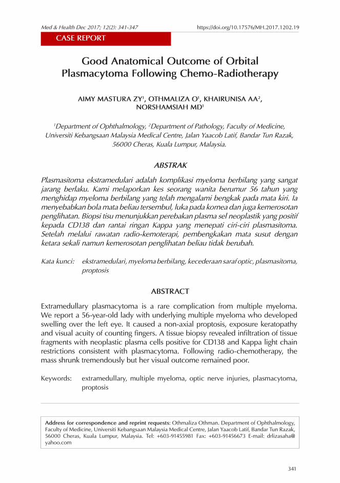
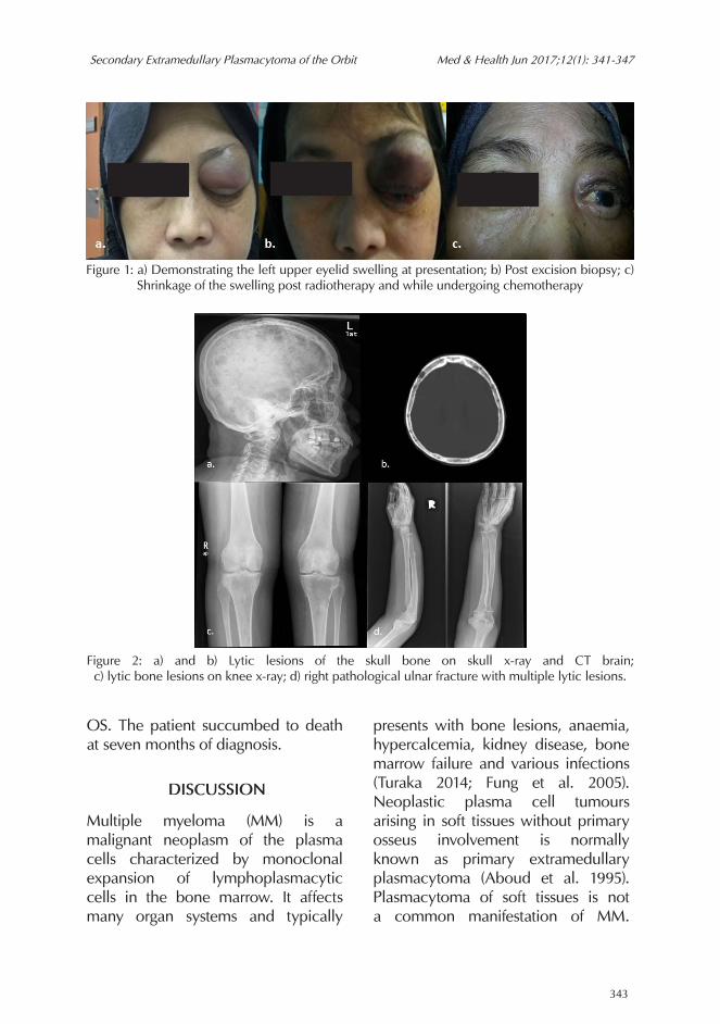
A 56-year-old Malay lady was diagnosed with multiple myeloma in April 2013 and received suboptimal treatment due to financial constraint. In 2015, she presented with a 3-wks history of left upper eyelid swelling. Within two weeks, the eyelid swelling doubled in size and was associated with proptosis, restriction of eye movement and poor vision in the left eye (Figure 1a). At the same time she was diagnosed with pathological right ulnar fracture with X-ray findings of multiple lytic lesion corresponding to myeloma bone disease (Figure 2a, 2c and 2d). She also had a previous history of pathological right humerus fracture one year prior to this presentation. Examinations revealed vision of counting finger OS (left eye). Relative Afferent Pupilary Defect was positive
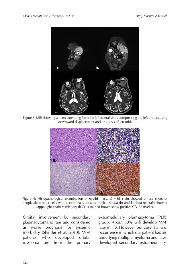
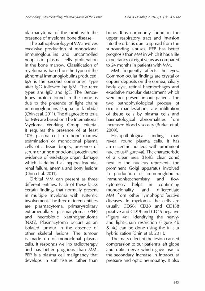
with restrictions of extraocular muscle movements in all gazes. There was a protruding mass on the upper eyelid which was non tender and was fixed to the overlying and underlying structure, causing a non-axial proptosis, hypotropia, lagophthalmos and a resultant exposure keratopathy (Figure 1a). The mass was firm on palpation and extended from the superior orbital rim towards the lid margin. Intraocular pressure was 24 mmHg OS and 11 mmHg OD (right eye). The left optic disc was swollen and hyperaemic; and vessels were tortuous and dilated. The mass effect caused choroidal folds around the arcades and macular striations. A computed tomography (CT) scan was done and it revealed a left orbital mass with local involvement and with the presence of a multiple lytic lesions of the skull vault (Figure 2b). Magnetic resonance imaging (MRI) showed a tissue mass superior to the globe extending from the left frontal sinus and compressing the globe and optic nerve downwards (Figure 3a-3d). There was no globe or optic nerve infiltration noted. A tissue biopsy revealed multiple tissue fragments infiltrated by neoplastic plasma cells which were positive for CD138 and Kappa light chain restriction suggestive of a secondary extramedullary plasmacytoma of the orbit (Figure 4). The patient completed 40 Gy/20 fragments of radiotherapy and is currently undergoing chemotherapy. The swelling improved tremendously after receiving the current treatment (Figure 1c). However, her vision remained poor with light perception
343
Secondary Extramedullary Plasmacytoma of the Orbit Med & Health Jun 2017;12(1): 341-347
OS. The patient succumbed to death at seven months of diagnosis.
DISCUSSION
Multiple myeloma (MM) is a malignant neoplasm of the plasma cells characterized by monoclonal expansion of lymphoplasmacytic cells in the bone marrow. It affects many organ systems and typically
presents with bone lesions, anaemia, hypercalcemia, kidney disease, bone marrow failure and various infections (Turaka 2014; Fung et al. 2005). Neoplastic plasma cell tumours arising in soft tissues without primary osseus involvement is normally known as primary extramedullary plasmacytoma (Aboud et al. 1995). Plasmacytoma of soft tissues is not a common manifestation of MM.
Figure 1: a) Demonstrating the left upper eyelid swelling at presentation; b) Post excision biopsy; c) Shrinkage of the swelling post radiotherapy and while undergoing chemotherapy
Figure 2: a) and b) Lytic lesions of the skull bone on skull x-ray and CT brain; c) lytic bone lesions on knee x-ray; d) right pathological ulnar fracture with multiple lytic lesions.
344
Med & Health Dec 2017;12(2): 341-347 Aimy Mastura Z.Y. et al.
Orbital involvement by secondary plasmacytoma is rare and considered as worse prognosis for systemic morbidity (Shinder et al. 2010). Most patients who developed orbital myeloma are from the primary
extramedullary plasmacytoma (PEP) group. About 50% will develop MM later in life. However, our case is a rare occurrence in which our patient has an underlying multiple myeloma and later developed secondary extramedullary
Figure 3: MRI showing a mass extending from the left frontal sinus compressing the left orbit causing downward displacement and proptosis of left orbit
Figure 4: Histopathological examination of eyelid mass. a) H&E stain showed diffuse sheet of neoplastic plasma cells with eccentrically located nuclei; Kappa (b) and lambda (c) stain showed
kappa light chain restriction; d) Cells stained brown show positive CD138 marker.
345
Secondary Extramedullary Plasmacytoma of the Orbit Med & Health Jun 2017;12(1): 341-347
plasmacytoma of the orbit with the presence of myeloma bone disease. The pathophysiology of MM involves excessive production of monoclonal immunoglobulins and uncontrolled neoplastic plasma cells proliferation in the bone marrow. Classification of myeloma is based on the type of the abnormal immunoglobulins produced. IgA is the second commonest type after IgG followed by IgM. The rarer types are IgD and IgE. The Bence-Jones protein found in the urine is due to the presence of light chains immunoglobulins (kappa or lambda) (Chin et al. 2011). The diagnostic criteria for MM are based on The International Myeloma Working Group criteria. It requires the presence of at least 10% plasma cells on bone marrow examination or monoclonal plasma cells of a tissue biopsy, presence of serum or urine monoclonal protein, and evidence of end-stage organ damage which is defined as hypercalcaemia, renal failure, anemia and bony lesions (Chin et al. 2011). Orbital MM can present as three different entities. Each of these lacks certain findings that normally present in multiple myeloma with systemic involvement. The three different entities are plasmacytoma, primary/solitary extramedullary plasmacytoma (PEP) and necrobiotic xanthogranuloma (NXG). Plasmacytoma occurs as an isolated tumour in the absence of other skeletal lesions. The tumour is made up of monoclonal plasma cells. It responds well to radiotherapy and has better prognosis than MM. PEP is a plasma cell malignancy that develops in soft tissues rather than
bone. It is commonly found in the upper respiratory tract and invasion into the orbit is due to spread from the surrounding sinuses. PEP has better prognosis than MM in which it has a life expectancy of eight years as compared to 24 months in patients with MM. MM frequently affects the eyes. Common ocular findings are crystal or copper deposits on the cornea, ciliary body cyst, retinal haemorrhages and exudative macular detachment which were not present in our patient. The two pathophysiological process of ocular manifestations are infiltration of tissue cells by plasma cells and haematological abnormalities from increased blood viscosity (Burkat et al. 2009). Histopathological findings may reveal round plasma cells. It has an eccentric nucleus with prominent nucleolus (Figure 4a). The characteristic of a clear area (Hoffa clear zone) next to the nucleus represents the prominent Golgi apparatus involved in production of immunoglobulin. Immunohistochemistry and flow cytometry helps in confirming monoclonality and differentiate MM from other lymphoproliferative diseases. In myeloma, the cells are usually CD56, CD38 and CD138 positive and CD19 and CD45 negative (Figure 4d). Identifying the heavy- and light-chain restriction (Figure 4b & 4c) can be done using the in situ hybridization (Chin et al. 2011). The mass effect of the lesion caused compression to our patient’s left globe and optic nerve which gave rise to the secondary increase in intraocular pressure and optic neuropathy. It also
346
Med & Health Dec 2017;12(2): 341-347 Aimy Mastura Z.Y. et al.
caused lagopthalmos and proptosis with subsequent exposure keratopathy.The myeloma bone disease is usually quite severe due to the multiple factors that contribute to the osteoclastic bone destruction and blocking of bone repair (Giuliani et al. 2004). Increase in osteoclastic activity is the primary cause of abnormal bone remodelling. This abnormal bone remodelling occurs in proximity to the active myeloma cells with a decrease in surrounding osteoblasts activity (Giuliani et al. 2004; Kristinsson et al. 2011). In contrast to myeloma bone disease, there are areas of bone without tumour involvement in myeloma patients which has balanced bone remodelling and an increased osteoclastogenesis combined with normal-to-increased bone formation. Permanent scarring of the bone can also be found in MM patients. Patients in remission may have persistent lytic lesions with no evidence of infiltration of the malignant myeloma cells in the bone marrow (Anderson et al. 2002). All these process contribute to lytic bone disease and causing pathological fracture in our patient. Reports stated that the interval to development of orbital lesions since the primary diagnosis of MM is an average of 18 months (Chin et al. 2011; Burkat et al. 2009). However, our patient developed EMP of the orbit approximately 31 months after diagnosis of MM with presence of myeloma bone disease and succumbed to death 7 months later. Radiotherapy has been proven to be effective in improving the symptoms by decreasing the size of lesions (Turaka
2014) as seen in our patient. The mass shrunk tremendously and improved the anatomical outcome of the patient despite her poor visual outcome. Treatment of the orbital lesions includes local excision as a salvage surgery, exenteration or radiotherapy and additional chemotherapy (Turaka 2014). In summary, our patient survived 38 months after the diagnosis of MM and only survived seven months after the diagnosis of secondary extramedullary plasmacytoma of the orbit. Though the radiotherapy and chemotherapy helped in improving anatomical outcome of our patient, she eventually died despite the aggressive treatment given.
CONCLUSION
Orbital MM depicts a more aggressive disease as compared to extraorbital plasmacytomas or primary/solitary extramedullary plasmacytoma (PEP).MM with orbital involvement is associated with poor prognostic outcome. Early detection may assist in early referral to the oncology/hematology team and may help in preventing the debilitating complications and thus help in improving the patient’s quality of life.
REFERENCESAboud, N., Sullivan, T., Whitehead, K. 1995. Primary
extramedullary plasmacytoma of the orbit. Aust N Z J Ophthalmol 23(3): 235–9.
Adkins, J.W., Shields, J.A., Shields, C.L., Eagle, R.C., Flanagan, J.C., Campanella, P.C. 1997. Plasmacytoma of the eye and orbit. Int Ophthalmol 20(6): 339–43.
Anderson, K.C., Shaughnessy, J.D., Barlogie, B., Harousseau, J.L., Roodman, G.D. 2002.
347
Secondary Extramedullary Plasmacytoma of the Orbit Med & Health Jun 2017;12(1): 341-347
Multiple myeloma. Hematology Am Soc Hematol Educ Program 385(9983): 214–40.
Burkat, C.N., Van Buren, J.J., Lucarelli, M.J. 2009. Characteristics of Orbital Multiple Myeloma: A Case Report and Literature Review. Surv Ophthalmol 54(6): 697–704.
Chin, K.J., Kempin, S., Milman, T., Finger, P.T. 2011. Ocular manifestations of multiple myeloma: Three cases and a review of the literature. Optometry 82(4): 224–30.
Fung. S., Selva, D., Leibovitch, I., Hsuan, J., Crompton, J. 2005. Ophthalmic manifestations of multiple myeloma. West Afr J Med 219(1): 43–8.
Giuliani, N., Colla, S., Rizzoli, V. 2004. New insight in the mechanism of osteoclast activation and formation in multiple myeloma: focus on the receptor activator of NF-kappaB ligand (RANKL). Exp Hematol 32(8): 685–91.
Kristinsson, S.Y., Minter, A.R., Korde, N., Tan, E., Landgren, O. 2011. Bone disease in multiple
myeloma and precursor disease: novel diagnostic approaches and implications on clinical management. Expert Rev Mol Diagno 11(6): 593–603.
Lida, N., Saito, K., Fukushima, K. 2005. A case of extramedullary plasmacytoma arising from the lacrimal gland: a case report. Eur J Plast Surg 28: 364–7.
Shinder, R., Al-Zubidi, N., Esmaeli, B. 2010. Survey of orbital tumors at a comprehensive cancer center in the United States. Head Neck 33(5): 610–14.
Turaka, A. 2014. Secondary Plasmacytoma of The Orbit In A Patient With Refractory Ig G Kappa Multiple Myeloma. Delhi J Ophthalmol 24(4): 245–7.
Received: 31 January 2017Accepted: 18 May 2017