Melioidosis in Pahang, Malaysia - e-mjm.org · PDF filemortality and outcome of adult...
8
I ORIGINAL.ARTICLE Melioidosis in Pahang, Malaysia 2i SH How, MMed*, K H Ng, MRCP***, AR Jamalludin, MPH (Epid & Biostat)**, A Shah, MSc. (Clin. Derm.)*, Y Rathor, MD* "Department of Internal Medicine, ** Department of Community Health and Family Medicine, Kulliyyah of Medicine, International Islamic University Malaysia, P.O.Box 141, 27510, Kuantan, Pahang, Malaysia, *'*Department of Internal Medicine, Hospital Tengku Ampuan Afzan, Kuantan Introduction' Melioidosis is caused by gram-negative bacilli, Burkholderia pseudomallei, which is a common soil and fresh water saprophyte especially in rice paddy fields in tropical and subtropical areas. Affected individuals usually have direct contact with soil as well as underlying predisposing factors. It frequently causes abscesses in almost all organs, but the commonest presentation is pneumonia!. It was associated with very high in-hospital mortality especially in the septicaemic form (65%) in the 1980s in Malaysia', In the past 20 years, several studies have shown that the mortality rate has been reduced to 19-37% with high dose ceftazidime, imipenem or cefoperazone-sulbactam therapy for at least two weeks!,3.4.5, Parenteral ammdcillin- clavulinic acid has been shown to give similar results but with higher treatment failure rates compared to ceftazidime 6 . The use of cotrimoxazole in addition to a beta-lactam is still controversiap,4, Pahang is one of 14 states in Malaysia. It has an area of 35,965 km' and a population of 1.28 million 038,000 were 18 years old in 2002 Malaysia Statistic Department). The main economic activity is agriculture, mainly rubber and oil palm cultivation. The incidence of melioidosis in this region is unknown, We conducted a retrospective study to determine the incidence, predisposing factors, clinical features, bacteriology, mortality and outcome of adult melioidosis in Pahang, Malaysia during the period from January 2000 to June 2003. Materials and Methods This is a retrospective study to identify cases of adult melioidosis (age above 18 years old) in Pahang during the period of January 2000 to June 2003. In Pahang, only Hospital Tengku Ampuan Afzan (HTAA) in Kuantan and Hospital Mentakab (HM) have facilities to This article was accepted: 2 June 2005 Corresponding Author: How Soon Hin, Kulliyyah of Medicine, International Islamic University Malaysia, P.D-Box 141,27510 Kuantan, Pahang, MalaYSia 606 Med J Malaysia Vol 60 No 5 December 2005
Transcript of Melioidosis in Pahang, Malaysia - e-mjm.org · PDF filemortality and outcome of adult...
I
ORIGINAL.ARTICLE
Melioidosis in Pahang, Malaysia
2i
S H How, MMed*, K H Ng, MRCP***, A R Jamalludin, MPH (Epid & Biostat)**, A Shah, MSc. (Clin.Derm.)*, Y Rathor, MD*
"Department of Internal Medicine, ** Department of Community Health and Family Medicine, Kulliyyah of Medicine, InternationalIslamic University Malaysia, P.O.Box 141, 27510, Kuantan, Pahang, Malaysia, *'*Department of Internal Medicine, Hospital TengkuAmpuan Afzan, Kuantan
Introduction'
Melioidosis is caused by gram-negative bacilli,Burkholderia pseudomallei, which is a common soiland fresh water saprophyte especially in rice paddyfields in tropical and subtropical areas. Affectedindividuals usually have direct contact with soil as wellas underlying predisposing factors. It frequently causesabscesses in almost all organs, but the commonestpresentation is pneumonia!. It was associated withvery high in-hospital mortality especially in thesepticaemic form (65%) in the 1980s in Malaysia', Inthe past 20 years, several studies have shown that themortality rate has been reduced to 19-37% with highdose ceftazidime, imipenem or cefoperazone-sulbactamtherapy for at least two weeks!,3.4.5, Parenteralammdcillin- clavulinic acid has been shown to givesimilar results but with higher treatment failure ratescompared to ceftazidime6. The use of cotrimoxazole inaddition to a beta-lactam is still controversiap,4,
Pahang is one of 14 states in Malaysia. It has an area of35,965 km' and a population of 1.28 million 038,000were 18 years old in 2002 Malaysia StatisticDepartment). The main economic activity is agriculture,mainly rubber and oil palm cultivation. The incidenceof melioidosis in this region is unknown, We conducteda retrospective study to determine the incidence,predisposing factors, clinical features, bacteriology,mortality and outcome of adult melioidosis in Pahang,Malaysia during the period from January 2000 to June2003.
Materials and Methods
This is a retrospective study to identify cases of adultmelioidosis (age above 18 years old) in Pahang duringthe period of January 2000 to June 2003. In Pahang,only Hospital Tengku Ampuan Afzan (HTAA) inKuantan and Hospital Mentakab (HM) have facilities to
This article was accepted: 2 June 2005Corresponding Author: How Soon Hin, Kulliyyah of Medicine, International Islamic University Malaysia, P.D-Box 141,27510Kuantan, Pahang, MalaYSia
606 Med J Malaysia Vol 60 No 5 December 2005
perform culture and sensitivity tests for B.pseudomallei. We therefore identified all cases withpositive culture for B. pseudomallei from blood, pus,cerebro-spinal fluid or other body fluids from thedatabase of the microbiology departments of these twohospitals within the study period. Case records ofpatients were reviewed and relevant data wasextracted, including demography, predisposing factors,clinical presentation, physical findings, laboratory andradiological investigations, antibiotic therapy andclinical outcome.
When a patient was found to have positive cultures forB. pseudomallei on more than one admission duringthe study period, the second and subsequent infectionswere considered as relapses. Patients with a culture~
proved infection before the study period wereconsidered to have a positive history of past infection.Appropriate antibiotic therapy was defined as antibiotictreatment with ceftazidime, cefoperazone-sulbactam,amoxicillin-clavulanic acid, imipenem or thecombination of any of these drugs. The in-hospitalmortality was calculated based on patients with aknown outcome (after excluding patients who weredischarged against the advice of doctors, transferred toanother hospital or had an uncertain outcome).
Both HTAA and HM used the same method for isolationof B. pseudomallei. Body fluids were cultured in bloodagar and MacConkey agar at 37°C and confirmation wasdone by observing the colony morphology, stainingreaction, motility and biochemical tests (indole, triplesugar iron, citrate, phenyl deaminase, oxidasefermentative 1% glucose, urease, oxidase and DNAse).Sensitivity testing was performed by agar dilutionmethod using BBL ™ Sensi-Disc™ AntimicrobialSusceptibility Test Discs (Becton, Dickinson andCompany, USA). Blood samples were incubated intryptic soy broth bottle (Roche) in BACTEC system andcultured in blood agar and MacConkey agar if positivegrowth was detected.
Statistical AnalysisThe data was analyzed using Statistical Package for theSocial Sciences (SPSS) version 10 software package.Incidence was calculated by. average number. of newcases per year divided by year 2002 adult population(;;,18 years old) in Pahang. Univariate analysis wasdone for all variables. Independent sample t-Test orMann-Whitney U Test was used for continuousparameters depending on their normality. Categoricalvariables were tested using chi-square test. Factorsfound to be significant on univariate analysis were then
Med J Malaysia Vol 60 No 5 December 2005
Melioidosis in Pahang, Malaysia
subjected to multivariate analysis using binary logisticregression. Significance was taken at 0.05.
Results
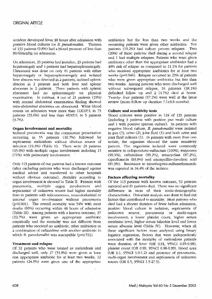
Cohort studyDuring the study period, a total of 157 patients hadpositive culture for B. pseudomallei from the two labsin HTAA and HM. Most of the cases presented at thetwo largest hospitals namely HTAA (44.4%) and HM(36.3%); the remaining cases were from other smallerdistrict hospitals. The calculated annual incidence ofadult melioidosis in Pahang state was 6.07 per 100, 000population per year. Only 135 patients' case recordscould be traced and analyzed. One hundred and sixpatients (78.5%) were male. There were 112 Malays(83%), 13 Chinese (9.6%), four Indians (3%), fouraborigines (3%) and two Indonesians. Most patientswere between 40 to 60 years of age and the medianwas 51 years (range: 19 - 87 years) (Table 1). Patientswithout predisposing factors were significantly younger(median age 41 years) than patients with predisposingfactors (median age 52 years) (p=0.012). Only sixpatients (4.4%) had a previous history of confirmedmelioidosis and another six patients (4.4%) hadprevious infection possibly due to melioidosis but withnegative cultures.
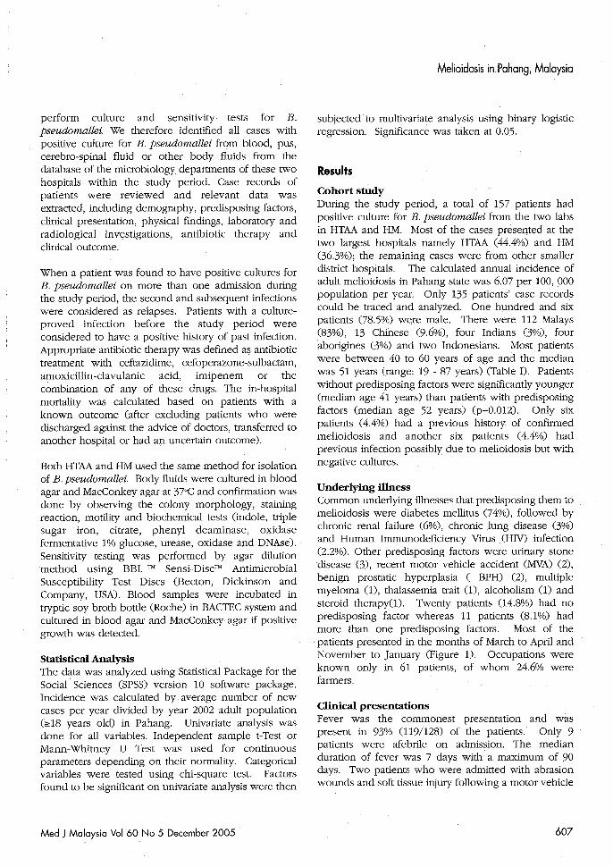
Underlying illnessCommon underlying illnesses that predisposing them tomelioidosis were diabetes mellitus (74%), followed bychronic renal failure (6%), chronic lung disease (3%)and Human Immunodeficiency Virus (HIV) infection(2.2%). Other predisposing factors were urinary stonedisease (3), recent motor vehicle accident (MVA) (2),benign prostatic hyperplasia ( BPH) (2), multiplemyeloma (1), thalassemia trait (1), alcoholism (1) andsteroid therapy(1). Twenty patients (14.8%) had nopredisposing factor whereas 11 patients (8.1%) hadmore than one predisposing factors. Most of thepatients presented in the months of March to April andNovember to January (Figure 1). Occupations wereknown only in 61 patients, of whom 24.6% werefarmers.
Clinical presentationsFever was the commonest presentation and waspresent in 93% (119/128) of the patients. Only 9patients were afebrile on admission. The medianduration of fever was 7 days with a maximum of 90days. Two patients who were admitted with abrasionwounds and soft tissue injury following a motor vehicle
607
ORIGINAL ARTICLE
accident developed fever 18 hours after admission withpositive blood cultures for B. pseudomallei. Thirteenof 133 patients (9.8%) had a blood pressure of less than90/60mmHg on admission.
On admission, 23 patients had jaundice, 23 patients hadhepatomegaly and 4 patients had hepatosplenomegaly.Ultrasound was done on only 11 patients with eitherhepatomegaly or hepatosplenomegaly and isolatedliver abscess was detected in 2 patients, isolated splenicabscess in 2 patients and both liver and splenicabscesses in 2 patients.. Three patients with splenicabscesses had no splenomegaly on physicalexamination. In contrast, 4 out of 21 patients (19%)with normal abdominal examination finding showedintra-abdominal abscesses on ultrasound. White bloodcounts on admission were more than l1X106/L in 75patients (55.6%) and less than 4X10⁶/L in 5 patients(3.7%).
Organ involvement and mortalityIsolated pneumonia was the commonest presentationoccurring in 55 patients (40.7%) followed bysepticaemic melioidosis without obvious source ofinfection 09.3%) (Table II). There were 21 patients05.6%) with multiple organ involvement including 15(71%) with pulmonary involvement.
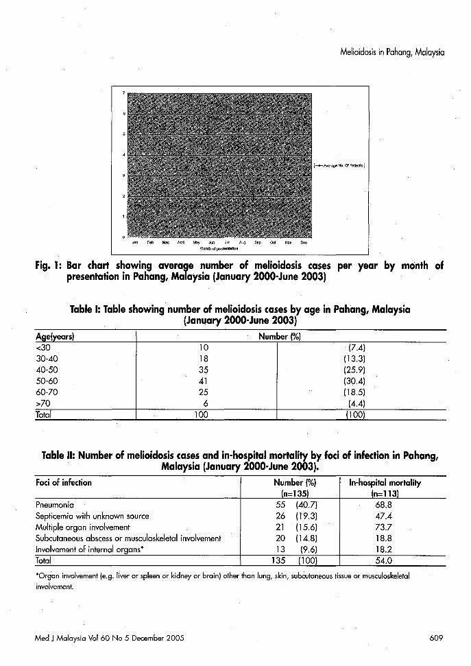
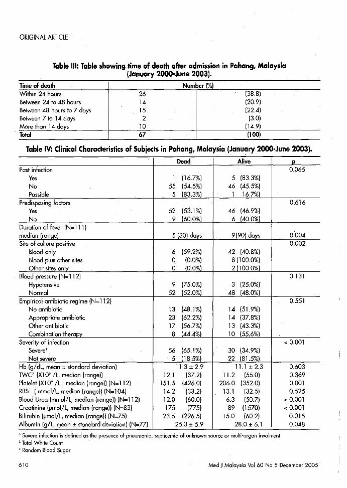
Only 113 patients of our patients had a known outcome(after excluding patients who were discharged againstmedical advice and transferred to other hospitalswithout obvious outcome). Mortality according toorgan involvement is showed'in Table II. Patients withpneumonia, multiple organ involvement andsepticaemic of unknown source had higher mortalitythan to patients with subcutaneous, musculoskeletal orinternal organ involvement without pneumonia(p<O.OOl). The overall mortality was 54% with mostdeaths (65%) occurring within 48 hours of admission(Table III). Among patients with a known outcome, 37(32.7%) were given an appropriate antibioticempirically and the mortality was not different frompatients who received no antibiotic, other antibiotics ora combination of ceftazidime with another antibiotic towhich B. pseudomallei was susceptible (p=0.55l).
Treatment and relapseOf 52 patients who were treated as melioidosis anddischarged well, only 27 (51.9%) were given at leastone appropriate antibiotic for at least two weeks, 12patients (24.5%) were given one of the appropriate
608
antibiotics but for less than two weeks and theremaining patients were given other antibiotics. Tenpatients 09.2%) had culture proven relapses. Two(20%) of these patients died during a second relapseand 1 had multiple relapses. Patients who were givenantibiotics other than the appropriate antibiotics had a40% risk of relapse as compared to 11.1% for patientswho received appropriate antibiotics for at least twoweeks (p=0.046). Relapse occurred in 25% of patientswho were given appropriate antibiotics but less thantwo weeks. Among patients who were discharged wellwithout subsequent relapse, 16 patients (38.1%)defaulted follow up and 2 (4.7%) died at home.Twenty- four patients (57.1%) were well at the latestreview (mean follow up duration 7.1±3.8 months).
Culture and sensitivity testsBlood cultures were positive in 124 of 135 patients(including 3 patients with positive pus swab cultureand 1 with positive sputum culture). In patients withnegative blood culture, B. pseudomallei were isolatedin pus (7), urine (2), joint fluid 0) and both urine andjoint fluid cultures 0). In patients with more .than oneisolate, the organism showed the same sensitivitypattern. The organisms isolated were commonlysensitive to cefoperazone-sulbactam (100%), imipenem(99.1%), ceftazidime (99.2%), tetracycline (87.2%),ciprofloxacin (83.9%) and amoxycillin-clavulinic acid(81.9%). Resistance to trimethoprim-sulfamethoxazolewas reported in 34.4% of the isolates.
Factors affecting mortalityOf the 113 patients with known outcome, 52 patientssurvived and 61 patients died. There was no significantdifference in term of their socio-demographiccharacteristics. Univariate analysis was done to find outfactors that contributed to mortality. Most patients whodied had a shorter duration of fever before admission,positive blood culture in isolation, septicaemia ofunknown source, pneumonia or multi-organinvolvement; a lower platelet count, higher serumcreatinine level, higher serum bilirubin level and lowerserum albumin level (Table IV). However, when allthese significant factors were analyzed using binarylogistics regression, factors that were independentlyassociated with the mortality of melioidosis patientswere duration of fever (OR 0.91, 95%CI 0.85-0.98),platelet count (OR 0.99, 95%CI 0.98-0.99), blood urea(OR 1.1, 95%CI 1.0-1.2) and presence of pneumonia,multi-organ involvement and septicaemia of unknownsource (OR 6.5, 95%CI 1.5-27.5).
Med J Malaysia Vol 60 No 5 December 2005
Melioidosis in. Pahang, Malaysia
i-.-Average No. Of Patients 1
~n ~ ~ ~ _ ~ ~ ~ ~p ~ ~ ~~
Month of presentation
Fig. 1: Bar chart showing average number of melioidosis cases per year by month ofpresentation in Pahang, Malaysia (January 2000-June 2003)
Table I: Table showing number of melioidosis cases by age in Pahang, Malaysia(January 2000-June 2003)
Age(years)<3030-4040-5050-6060-70>70Total
10183541256
100
Number (%)(7.4)
(13.3)(25.9)(30.4)(18.5)(4.4)
(l00)
Table II: Number of melioidosis cases and in-hospital mortality by foci of infection in Pahang,Malaysia (January 2000-June 2003).
Foci of infection Number (%) In-hospital mortality(n=135) (n=113)
Pneumonia 55 (40.7) 68.8Septicemia with unknown source 26 (19.3) 47.4Multiple organ involvement 21 (15.6) 73.7Subcutaneous abscess or musculoskeletal involvement 20 (14.8) 18.8Involvement of internal organs' 13 (9.6) 18.2Total 135 (l00) 54.0
'Organ involvement (e.g. liver or spleen or kidney or brain) other than lung, skin, subcutaneous tissue or musculoskeletal
involvement.
Med JMalaysia Vol 60 No 5 December 2005 609
ORIGINAL ARTICLE
Table III: Table showing time of death after admission in Pahang, Malaysia(January 2000·June 2003).
Time of deathWithin 24 hoursBetween 24 to 48 hoursBetween 48 hours to 7 daysBetween 7 to 14 daysMore than 14 days
. Total
2614152
1067
Number (%)(38.8)(20.9)(22.4)
(3.0)(14.9)(100)
Table IV: Clinical Characteristics of Subjects in Pahang, Malaysia (January 2000·June 2003).
Dead Alive p
Past infection 0.065Yes 1 (16.7%) 5 (83.3%)No 55 (54.5%) 46 (45.5%)possible 5 (83.3%) 1 16.7%)
Predisposing factors 0.616Yes 52 (53.1%) 46 (46.9%)No 9 (60.0%) 6 (40.0%)
.Duration of fever (N=111 )median (range) 5 (30) days 9(90) days 0.004Site of culture positive 0.002
Blood only 6 (59.2%) 42 (40.8%)Blood plus other sites 0 (0.0%) 8 (100.0%)Other sites only 0 10.0%1 21100.0%1
Blood pressure (N=112) 0.131Hypotensive 9 (75.0%) 3 (25.0%)Normal 52 (52.0%) 48 (48.0%)
Empirical antibiotic regime (N=112) 0.551No antibiotic 13 (48.1%) 14 (51.9%)Appropriate antibiotic 23 (62.2%) 14 (37.8%)Other antibiotic 17 (56.7%) 13 (43.3%)Combination therapy 8 (44.4%1 10 (55.6%1
Severity of infection < 0.001Severel 56 (65.1%) 30 (34.9%)Not severe 5 (18.5%) 22 (81.5%1
Hb (g/dL, mean ± standard deviation) 11.3±2.9 11.1 ± 2.3 0.603TWe (Xl 09 IL, median (range)) 12.1 (37.2) 11.2 (55.0) 0.369Platelet (Xl 09 IL , median (range)) (N=112) 151.5 (426.0) 206.0 (352.0) 0.001RBS3 (mmol/L, median (range)) (N=104) 14.2 (33.2) 13.1 (32.5) 0.525Blood Urea (mmol/L, median (range)) (N=112) 12.0 (60.0) 6.3 (50.7) < 0.001Creatinine (IJmol/L, median (range)) (N=83) 175 (775) 89 (1570) < 0.001Bilirubin (IJmol/L, median (range)) (N=75) 23.5 (296.5) 15.0 (60.2) 0.015Albumin (giL, mean ± standard deviation) (N=77) 25.3 ± 5.9 28.0 ± 6.1 0.048
1 Severe infection is defined as the presence of pneumonia, septicemia of unknown source or multi-organ involment2 Total White Count3 Random Blood Sugar
610 Med J Malaysia Vol 60 No 5 December 2005
Discussion
The incidence of melioidosis varies between countriesand also between different parts of the same country.In Thailand, it is most commonly seen in the northernregion and the incidence was 3.6-5.5 per 100,000 peryear7. In northern Australia, the incidence was 16.5 per100,000 per year 3. We are not aware of any publisheddata on the incidence of melioidosis in Malaysia. Webelieve the incidence may vary among different statesas agricultural activities differ in the various states. Theincidence of 6.07 per 100,000 per year recorded in ourstudy is comparable with Thailand. This may representthe incidence of melioidosis in most states in Malaysia.However, the incidence may be lower in states withlimited agricultural activities especially in Kuala Lumpuror Penang. The picture seen in these two states maybe similar to the incidence reported in Singapore (1.7per 100,000 per year) 8. The true incidence may behigher than reported as some patients may havenegative cultures despite extensive investigation. In ourstudy, there were 6 patients who had possible pastinfection. Three of these patients had a history ofsubcutaneous abscesses, one had pneumonia whichresponded to ceftazidime, one had septic arthritis andanother had multiple liver abscesses with positive'culture only during the subsequent admission.
The majority of our patients were Malay (83%). Thismay be because more Malays in Pahang work in theagricultural sector. The male to female ratio of 3.6 to 1reflects the importance of occupational exposure and isconsistent with a study done in Northern Australia3.Occupations were recorded for 45% of our patients anda history of soil contact among non-farmers was notroutinely sought by the doctor. This makes analysis ofsoil contact as a risk factor difficult. The median age of51 years in our study was slightly higher than in otherreports 3,7 due to the exclusion ofpaediatric cases.
Diabetic mellitus is the most commonly describedpredisposing factor in the literature. To ourknowledge, the incidence of diabetic patients (74%) inour study is the highest reported (37% to 60%)'·2.3. Thiscould be due to the higher prevalence of diabetes inMalaysia (more than 8%) as compared to Caucasians inAustralia (5% to 6%) 10 and poor diabetic control inmost of the diabetic patients". Poor diabetic control isan important risk factor for melioidosis9 • Bothalcoholism and chronic lung disease were relativelyuncommon in our study compared to NorthernAustralia3 as most of our patients were Muslims andalcohol is prohibited in Islam. The incidence of cystic
Med J Malaysia Vol 60 No 5 December 2005
Melioidosis in Pahang, Malaysia
fibrosis is also low in Malaysia. We had three patientswho were HIV serology positive without otherpredisposing factors which has not been reportedearlier. BPH is known to cause urinary stasis andurinary tract infection but there was no publish data onBPH as a predisposing factor for melioidosis. We foundtwo patients with BPH presenting with fever anddysuria. Both of their urinalysis suggestive of urinarytrait infection and blood cultures were positive for B.pseudomallei. One of these patients also had diabetesmellitus and chronic renal failure. Unfortunately, urinecultures were not sent for these patients. Further studyneed to be done to look for possible associationbetween BPH and melioidosis.
In this study, patients without predisposing factorswere Significantly younger than patients withpredisposing factors. This could be related toundetected underlying predisposing factors such asthalassemia trait. In contrast, diabetes occurs morecommonly in older patients and may be more easilydetected.
Trauma to the skin due to motor vehicle accidents maybe an important predisposing factor as contact of thewound with soil may result in direct inoculation of theorganisms into the blood stream as seen in ,two of ourpatients. However, wound swabs were not taken fromeither patient to confirm our hypothesis. We concludedthat melioidosis should be considered in immunocompromised patients who develop fever after MVA inendemic areas and ceftazidime should be considered ifthe fever is not responding to the usual antibiotic.
In our study, 19% of our patients with normalabdominal examination were found to have intraabdominal abscesses on ultrasound. This reflects thelow sensitivity of physical examination in localizationof intra-abdominal abscesses in melioidosis. In ourstudy and most studies in Thailand, 1. 7 prostaticabscesses were not commonly encountered ascompared to 18% in Northern Australia3. This isprobably due to under diagnosis as most physicianshere do not perform CT scan routinely to locate intraabdominal abscesses in confirmed cases. Therefore, westrongly suggest routine radiological investigation andevaluation of the sensitivity of ultrasound as comparedto CT scan.
The mortality of melioidosis is extremely highespecially in the bacteraemic form. The reportedincidence of the bacteraemic form ranges from 46% to
611
ORIGINAL ARTICLE
60% 3, 7 as compared to 91.8% in our study, The higherincidence in our study may due to late presentation ofour patients who usually seek traditional treatmentbefore presenting to us, Higher frequency of diabetesmellitus in our study may be another contributing factoras diabetics are more at risk for bacteraemicmelioidosis9, Furthermore, paediatric patients whocommonly present with localized melioidosis wereexcluded in this analysis" (we identified 14 paediatriccases and this is reported separately),
The overall in-hospital mortality (53.5%) in this study ishigher than in other recent studies (19% to 37%) 3.4.5,7 asonly 51.9% of patients received an appropriateantibiotic regime despite positive culture, This isprobably due to lack of awareness among doctorsregarding the treatment of melioidosis as reported fromSouth India where the mortality was 58%13,Furthermor~, there were also more bacteraemicpatients in our study as compared to other studies 3, 7,
We also found a higher risk of relapse followinginappropriate antibiotic therapy which may result inmortality during relapse as seen in Thailand 14, In viewof the high mortality in septicaemic melioidosis, furtherstudies should be carried out to evaluate possibleapproaches in reducing the mortality rate, To date,there is no evidence that early treatment of suspected
1. White NJ. Melioidosis, Lancet 2003; 361: 1715-22,
2, Puthucheary SD, Parasakthi N, Lee MK. Septicaenicmelioidosis: a review of 50 cases from Malaysia, Trans RSoc Med Hyg 1992; 86: 683-85,
3, Currie B], Fisher DA, Howard DM, et aL Endemicmelioidosis in tropical northern Australia: a 10 yearprospective study and review of the literature, Clin InfectDis 2000; 31: 981-86,
4, Samuel M, Ti TY, Interventions for treating melioidosis,The Cochrane Database of Systematic Reviews, 2003,
5, White N], Dance DA, Chaowagul W, et aL Halving of
612
cases with empirical ceftazidime may reduce mortality,In our study, empirical ceftazidime or other appropriateantibiotic appeared to have no effect on mortality eventhough combination therapy was associated with abetter outcome, Further randomized studies arenecessary to resolve this matter, The use of G-CSF insepticaemic melioidosis needs further evaluation, 15
Whether early diagnosis using test kits16, serology,monoclonal antibody'7 and polymerase chain reaction(PCR) 18 will reduce the mortality rate requires furtherresearch,
In conclusion, melioidosis in Pahang is commonlyassociated with diabetes mellitus and mortality remainsextremely high despite antibiotic therapy,
Acknowledgements
We would like to acknowledge the assistance of thefollowing: Dr K Ramachandram, Mr Alex Francis(MSc), Puan Marsitah bt Abdul Jalil, Encik Zulkiflj b,Muda, Microbiology Department, Hospital TengkuAmpuan Afzan,
We are grateful for financial support from InternationalIslamic University Malaysia Research Centre,
mortality of severe melioidosis by ceftazidime, Lancet1989; 2: 697-701.
6, Suputtamongkol Y, Rajchanuwong A, Chaowagul W, et aLCeftazidime vs, amoxicillin/clavulanate in the treatmentof severe )1lelioidosis, Clin Infect Dis 1994; 19: 846-53,
7, Suputtamonkol Y, Hall A], Dance DAB, et aL Theepidemiology of melioidosis in Urbon Ratchatani,northen eastern Thalland, Int] Epidemiol 1994; 23: 108289,
8, Heng BH, Goh KT, Yap EH, et aL Epidemiologicalsurveillance of melioidosis in Singapore, Ann Acad MedSingapore 1998; 27: 478-84,
Med J Malaysia Vol 60 No 5 December 2005
9. Jenney AW, Lum G, Fisher DA, et al. Antibioticsusceptibility of Burkholderia pseudomallei from tropicalnorthern Australia and implications for therapy ofmelioidosis. Int J Antimicrob Agents 2001; 17: 109-13.
10. Cockram CS. The epidemiology of diabetes mellitus inthe Asia-Pacific region. HKMJ 2000; 6: 43-52.
11. Merriman A, Ross 1. Finding among 100 Type 2 diabeticsin a clinic in Penang, Malaysia, 1983-84. Ann Acad MedSingapore 1985; 14: 277-85.
12. Pagakrong L, Surapon V. Clinical manifestations ofmelioidosis inchildren. Paediatric Infect Dis J 1995; 14:136-40.
13. Jesudason MV, Anbarasu A, John T]. Septicaemicmelioidosis in a tertiary care hospital in south India.Indian J Med Res 2003; 117: 119-21.
14. Chaowagul W, Suputtamongkol Y, Dance DA, et al.Relapse in melioidosis: incidence and risk factors. J Infect.Dis 1993; 168: 1181-5.
Med J Malaysia Vol 60 No 5 December 2005
Melioidosis in Pahang, Malaysia
15. Nelson S, Belknap SM, Carlson RW, et al. A randomizedcontrolled trial of filgrastim as an adjunct to antibiotics fortreatment of hospitalized patients with communityacquired pneumonia. CAP Study Group. J Infect Dis 1998;178: 1075-80.
16. Wuthiekanun V, Amornchai P, Chierakul W, et al.Evaluation of immunoglobulin M (IgM) and IgG rapidcassette test kits for diagnosis of melioidosis in an area ofendemicity. J Clin Microbiol 2004; 42: 3435-7.
17. Dharakul T, Songsivilai S, Smithikarn S, et al. A Rapididentification of Burkholderia pseudomallei in bloodcultures by latex agglutination using lipopolysaccharidespecific monoclonal antibody. Am J Trop Med Hyg 1999;61: 658-62.
18. Dharakul T, Songsivilai S, Viriyachitra S, et al. Detectionof Burkholderia pseudomallei DNA in patients withsepticemic melioidosis. J Clin Microbiol 1996;.34: 609-14.
613