SCREENING AND CHARACTERIZATION OF FIBROLYTIC … and Characterization of... · morfologi, penilaian...
24
• I I * SCREENING AND CHARACTERIZATION OF FIBROLYTIC BACTERIA FROM SAGO WASTE Mohd Almuhsen Bin Karim QD 311 M697 1011 Bachelor of Science with Honours (Resource Biotechnology) 2012
-
Upload
dinhkhuong -
Category
Documents
-
view
227 -
download
0
Transcript of SCREENING AND CHARACTERIZATION OF FIBROLYTIC … and Characterization of... · morfologi, penilaian...
• I I *
SCREENING AND CHARACTERIZATION OF FIBROLYTIC BACTERIA FROM SAGO WASTE
Mohd Almuhsen Bin Karim
QD 311 M697 1011 Bachelor of Science with Honours
(Resource Biotechnology) 2012
--... ----~- - - ~--
Pusat Khidmat Maklumat Akadf'mik VNlVERSm MALAYSIA SARA\\AK
Screening and Characterization of Fibrolytic Bacteria From Sago Waste
Mohd Almuhsen bin Karim (24062)
This project is submitted in partial fulfillment of the requirement of the degree of Bachelor of Science with Honours
(Resource Biotechnology)
Supervisor: Dr Awang Ahmad Sallehin Bin Awang Husaini Co-supervisor: Dr. Azham Zulkharnain
Resource Biotechnology Department of Molecular Biology
Faculty of Resource Science and Technology University Malaysia Sarawak
2012
" I '
' . ' I ,
Acknowledgements
Bismillahirrahmanirrahim
Alhamdulillah. Praise to Allah that I have finished my Final Year Project. I am blessed
with Allah's supreme affection making me stronger day by day facing the difficulties and
constrains in finishing this project.
I would like to thank and express my deepest appreciation to my supervisor, Assoc.
Prof Dr. Awang Ahmad Sallehin Bin Awang Husaini for his supervision, helps and guidance
throughout my works in finishing this project.
I also would like to thank to my dear parents and family for their endless supports all
the time especially financial support and motivation support to me. They are the strength that
keeps me motivated in facing all the obstacles in completing this project.
In addition, I would like to express my sincere gratitude to postgraduate students of the
Molecular Genetic Laboratory, Microbiology Laboratory and Virology Laboratory of
UNIMAS, for their kindness in giving assistance in this project. Especially, to Christy Chan
Sien Wei who generously giving advice and guidance, also spending time to help me in
resolving my problems during finishing this project.
Also, thank you very much to the special peoples behind me all the time in UNIMAS,
my close friends who always accompany me when I am alone, encouraging me, listen to my
problem, supporting me and being a wonderful friends to me. Lastly, to my course mates and
labmates who kindly sharing, helping and caring during the course of this project.
.,. Punt Khidmat Maklumat Akademik UNlVERSm MALAYSIA SARAWAK
Table of Contents
Acknowledgement Table of Contents 11
List of Abbreviations iii List of Tables and Figures IV
Abstract I 1.0 Introduction 2 2.0 Literature Review 3
2.1 Sago waste 3 2.2 Fibrolytic bacteria 4 2.3 Cellulase and xylanase 5 2.4 Screening and characterization 6
3.0 Materials and Method 7 3.1 Sample 7 3.2 Media preparation 7
3.2.1 Xylan containing agar 7 3.2.2 CMC Agar 7 3.2.3 Enrichment medium 8 3.2.4 Slant agar 8
3.3 Enrichment and isolation of bacteria 8 3.4 Secondary Screening 9 3.5 Morphological characterization 9
3.5.1 Gram Staining 9 3.6 Biochemical test 10 3.7 Enzyme assays 10
3.7.1 Cellulase activity 10 3.7.2 Xylanase activity 10
3.8 Preservation of isolates 11 3.9 16s rONA analysis II
3.9.1 Genomic DNA Extraction II 3.9.2 Polymerase Chain Reaction II 3.9.3 Agarose Gel Electrophoresis 12
4.0 Results and Discussion 13 4.1 Primary screening and isolation 13 4.2 Secondary screening 14 4.3 Gram staining and morphological observation 18 4.4 Biochemical tests 19 4.5 Genomic DNA extraction 21 4.6 PCR 22 4.7 Enzyme analysis (cellulase and xylanase) 27
5.0 Conclusion and recommendation 30 References 31 Appendices 33
ii
" ',.· t. , f: t"; "
List of Abbreviation
CMC Carboxymethylcellulase
MR test Methyl Red
VP Test Voge-Proskauer Test
DNS 3,5-dinitrosalicylic acid
AGE Agarose Gel Electrophoresis
PCR Polymerase Chain Reaction
DNA Deoxyribonucleic Acid
ml Mililitre
III Microlitre
M Molar
roM Milimolar
run nanometer
O.D Optical Density
°c Degree Celcius
iii
-~-- - -~
.. .,'
List of Tables and Figures
Table Page
Table 3.1 Programme for PCR 12
Table 4.1 Samples, numbers and isolates names. 14
Table 4.2 Production of halos on the com cobs agar plates. 17
Table 4.3 Production of halos on the CMC agar plates. 17
Table 4.4 Biochemical tests results 19
Table 4.5 BLAST result for A2 sample 24
Table 4.6 BLAST result for A4 sample 24
Table 4.7 BLAST result for B 1 sample 25
Table 4.8 BLAST result for D2 sample 25
Figure Page
Figure 4.1 Halo produced on the CMC agar 15
Figure 4.2 Halo produce on 1 % com cobs agar 16
Figure 4.3 Genomic DNA bands for bacterial isolates A2, A4, Bl, D2 21
Figure 4.4 PCR products of A2, A4, Bl and D2. 23
Figure 4.5 The graft ofxylanase enzyme activity against time. 27
Figure 4.6 The graft of cellulase enzyme activity against time. 28
iv
Screening and Characterization of Fibrolytic Bacteria from Sago Waste
Mohd Almuhsen bin Karim
Resource Biotechnology Programme Oepatment of Molecular Biology
Faculty of Resource Science and Technology UNlMAS
Abstract
A total of 16 bacteria were successfully isolated from the sago waste after selectively enriched in media containing 1 % (w/v) of corn cobs. Screening of the bacterial isolates was done by using Congo-red staining method after the bacterial strains was cultured onto the CMC agar plates and 1 % corn cobs agar plates, respectively. Four isolates were chosen and selected for further analysis due to their ability to produce large halo and characterized. Standard biochemical test, morphological characterization, cellulase enzyme assay, xylanase enzyme assay and molecular characterization were done on these four isolates. Enzyme assays were conducted using ONS method. After ONA sequencing with 16s rONA analysis, the isolates were found to be similar to Bacillus pumilus and Bacillus subtiUs.
Key words: fibrolytic bacteria, biochemical test, sago waste, screening, characterizing, enzyme, 16s rONA.
Abstak
Sejumlah 16 bakteria telah berjaya dipencilkan dari sisa sagu setelah diperkayakan dalam media yang mengandungi 1% sisa tongkol jagung. Pemilhan bakteria telah dilakukan dengan menggunakan pewarnaan Congo-red setelah dikulturkan di atas agar CMC and agar 1% sisa tongkol jagung, masing-masing. Empat bakteria dipilih untuk ujikai selanjutnya kerana keupayaan untuk menghasilkan zon halo dan telah dikenalpasli. Ujian piawai biokimia, morfologi, penilaian enzim cellulase, penilaian enzim xylanse dan kajian ke atas ciri-ciri molekul lelah dilakukan ke atas bakteria ini. Penilaian enzim telah dijalankan menggunakan kaedah DNS. Selepas urutan DNA dikaji menggunakan 16s rDNA, isolat memiliki persamaan urutan DNA dengan Bacillus pumilus_dan Bacillus subtilis~
Kala kunci: bakleria fibrolitik, ujian biokimia, sisa sagu, pemilihan, kajian ciri, enzim, 16s rDNA
1
,, ', . • j . I ( I ,r. .'
1.0 Introduction
Industries that used plant as their sources usually produced a vast amount of residue into the
environment including sago starch processing industries. The major difficulties for a bigger
sago starch processing factory is that, the large amount of fibrous waste containing starch that
they produced, (Awg-Adeni et al., 2009). According to Shiroth et al., (1999), high amount of
starch contents and moisture present in this waste making it hard to manage. Hence, this waste
eventually caused pollution to environment. In fact, Abd-Aziz (2002) stated that cellulosic
fibrous content in starch residue is a very strong pollution. Thus, managing this waste is
crucial in order to reduce pollution and contribute to a sustainable development.
In addition, the product from managing this waste could be a market valuable product.
The starch presents in the fibrous sago waste can be hydrolyzed into useful glucose (pei-Lang
et al., 2006) to be used as low cost nutrient source in fennentation processes for the
biotechnology industry. Perhaps, apart from resolving the problem of pollution, exploitation
of sago waste will also giving an economic solution to manage the residue at sago processing
mills, (Awg-Adeni et al. 2009). Plus, agro-residue from sago starch processing industries is
abundantly, cheap and readily available especially in the state of Sarawak, Malaysia, (Apun et
al.. 2000).
Using bacteria as one of the agents in bioconversion of this sago waste to value added
end product is promising. Bacteria that are capable of converting sago waste by producing
extracellular enzymes that degrade the cellulosic waste to fennentable sugars . Specifically,
fibre that are contained inside the sago waste is degraded by fibrolytic bacteria in which, these
2
bacteria produces extracellular enzymes which are cellulase and xylanase. These enzymes are
valuable to the industries. Recently, xylanase has increased the interest of the researchers
because of its various applications in agro-industrial processes such as conversion of biomass
into fuels and chemicals, animal feedstock and many more.
The main objectives of this project were therefore to screen and characterize fibrolytic
bacteria from sago waste. In order to achieve this main objective:
I' 1. The morphological examination and biochemical characterization were performed on the
fibrolytic bacteria isolated.
11. Enzyme assays were conducted on the bacteria isolated.
iii. Genotypic characterization was done by using I6s rDNA for molecular characterization.
2.0 Literature Review
2.1 Sago waste
According to Awg-Adeni et ai., (2009), residue is a substance resulting from the processing of
a product. This residue can be co-product or by-product when profitable use is made from it.
If not, it is called waste which is termed as a material with no apparent market, social or
environmental value that caused pol1ution to environment.
Sago 'hampas' is the solid residue from the sago starch processing activity, (Awg
Adeni et ai., 2009). It is fibrous pith residue from crushing and sieving to get starch to
produce starch-rich flour. It contain mostly starch and lignocellulosic. Starch and
lignocellulosic is a good material as substrate for solid substrate fermentation . The 'hampas'
may be used as animal feed, compost for mushroom culture, for hydrolysis to confectioners'
3
.• ' f ~ . ~ ;. "
syrup and for particleboard manufacture (Phang et aI., 2000). The sago 'hampas' largely
consisted of cellulose and lignins that have some potential as a biosorbent (Vickineswary et
al., 1994). The study by Apun et al. (2000) identify Bacillus amyloliquefaciens UMAS 1002
from sago waste in which this bacteriwn has the ability to hydrolyze sago 'hampas' into
reducing sugars. From the studies amylolytic and cellulolytic enzymes also being produced by
the bacteria, thus create an extra advantage in the degradation of sago'hampas' .
2.2 Fibrolytic Bacteria
Fiber degrading bacteria always associated with the cellulolytic and hemicellulolytic bacteria.
Bacterial composition will adapt to the supply of high levels of dietary fiber by increased
growth of bacteria with cellulolytic and hemicellulolytic activities ( Varel et al., 1987; Durnic
et al.. 1998; Lesser et al.. 2000 as cited in Metzler-Zebeli, 2007).
According to Schmidt (2009), cellulolytic bacteria can be distinguished as primary
cellulolytic bacteria and as facultative cellulolytic bacteria. The primary cellulolytic bacteria
specialized in cellulose degradation to such an extent that they are unable to grow if this
compound is not present (Cytophaga. Sperocytophaga and Sporangium), while the facultative
cellulolytic bacteria are able to utilize organic compound (Vibrio Cellxibrio, and
Cellfalcicula). Cellulolytic bacteria are quite common and have capacity to break down lignin
and other wood components such as resins, gums, dye, tanic acid, waxes, and fats. In this
project, cellulolytic bacteria that responsible to degrade the sago palm into sago waste were
identified.
4
..
Pusat Khidmat Maklumat Akadfmik VNlVERSm MALAYSIA SARAWAK
On the other hand, the major class of hemicellulose is xylan, which is found in large
quantites in annual plants and deciduous trees, and in smaller quantities in conifers (Johri et
ai., 1999). Xylanase is an enzyme that can degrade xylan into simple sugars (Arora and
Bhatnagar., 2005). Hence, xylanolytic bacteria could degrade xylan into simple sugars.
2.3 Cellulase and Xylanase
Xylan is a complex polysaccharide having a backbone of xylose residues linked by I, 4
glycosidic bonds. It constitutes the major hemicellulose component of the plant cell. The main
chain of xylan consists of xylopyranose residues. Most xylans contain different substituent
groups such as acetyl, arabinosyl, and glucuronysyl residues in their backbone and side chains
(Beg et at., 200 I). Many reports on thermostable xylan digesting Bacillus species, Bacillus
amyloliquefaciens (Breccia et ai., 1998), BaciIrus sp. strain SPS-O (Bataillon et ai.,
2000) are available. Alicyclobacillus acidocaidarius (ATCC 27009) has been reported to
produce extracellular thermoacidophilic xylanase (Eckert et ai., 2002). Xylanases are
biotechnologically important enzymes since the hydrolysis products of xylans are used as
thickeners or as fat substitutes in food industry. Xylose and xylooligosaccharides are also used
to obtain liquid fuel, single cell proteins, and solvents (Eckert et ai., 2002). Xylanolytic
enzymes are also used in extraction and clarification of juices and wines, modification of
cereal flours to enhance the volume, textural and staling properties of bread, prebleaching of
paper pulps, retting of flax, hemp, and jute (Gilbert and Hazlewood, 1993).
Cell uloses are complex and heterogeneous polymer formed of D-glucose residues
linked by I, 4-glycosidic bonds. Cellulases are classified into three groups: exoglucanases,
5
' . f f ,. '
endoglucanases and D-glucosidases. Exoglucanases cleave the cellobiosyl units from the
nonreducing ends of the cellulose chains. Endoglucanases hydrolyse the internal cellulosic
linkages and D-glucosidases specifically cleave glucosyl units from the nonreducing ends of
cellooligosaccharides (Schiilein et ai., 2000). Cellulases are widely used in textile industry for
bio-polishing of fabrics, house hold laundry detergents, animal feeds, fruit juice processing,
baking and in de-inking of paper (Mavadza et aI., 2000).
2.3 Screening and characterization
Biochemical test is a part of the crucial method in screening and characterizing
bacteria. Biochemical test can classify bacteria into genus, subspecies or genus. This is
because each species of bacterium has a different molecule of DNA. Since DNA encodes for
protein synthesis, then different species of bacteria wilJ synthesize different protein enzymes.
Enzymes catalyze all the various chemical reaction of which the organism is capable. This in
turn means that different species of bacteria must carry out different sets of biochemical test.
Several standard biochemical tests are done in this project in order to screen and characterize
the fibrolytic bacteria. Apart from that, morphology identification also contribute in
characterizing the bacteria such as its shape, spore formation, cell wall etc. Another crucial
step yet precise is by using the PCR based method which require to analyze the DNA of the
bacteria. This method can distinguish and differentiate bacteria at the molecular level.
3.0 MATERIALS AND METHODS
3.1 Sampling
Samples consisted of sago waste were taken from Ngitsei sago factory from the division of
Mukah, state of Sarawak, Malaysia. The four different sago waste samples were used in which
designed as A, B, C and D.
3.2 Media Preparation
3.2.1 Xylan containing agar
Xylan medium was prepared for the growth of the microorganism. For each Petri dish, 15 cm3
ofxylan containing agar and the medium contained the following in giL of 0.1% yeast extract,
1 % xylan from com cob, 2% agar, 0.4% KH2P04, 0.2% NaC!, 0.1 % MgS04.7H20 and 0.005%
MnS04.H20 at pH7.0, Nanmori et. aI., (1990).
3.2.2 CMC Agar
CMC agar is used in screening procedure. The medium used consisted of the following in giL
of 0.2% of yeast extract, 0.1 % of KH2P04, 0.1 MgS04 and 0.5% of CMC.
7
3.2.3 Enrichment medium
Selective enrichment medium was prepared in which specifically com cobs that serve as the
sole carbon source and nutrient in the mediwn. Enrichment broth was prepared as stated in
Section 3.2.1 and excluding the agar.
3.2.4 Slant agar
Slant agar was prepared for a longer time storage as compared to the growth of bacteria on the
plate agar. The slant agar is prepared using the same composition as stated in Section 3.2.1.
3.3 Enrichment and isolation of bacteria
Selective enrichment of bacteria was done in order to enrich the amount of desired bacteria
and at the same time to suppress the unwanted bacteria. Mediwn that used in this selective
enrichment acted as primary selective media. One gram of each samples were put inside the
100 mL conical flask containing 100 mL of enrichment broth. Each flask was incubated for 24
hours III the incubator shaker for 200rpm at the temperature of 37°C.
Isolation of bacteria is crucial to get the pure culture involves direct plating method.
This also includes serial dilution method. The steps were modified from Nurulnisa (2009).
One hundred microliter of samples from the enrichment broth was added into 900 ilL of saline
water inside the eppendorftube. The sample was mixed together. Then, lOO ilL of the sample
was pipetted aseptically into the lO-1 dilution tube. The dilution was mixed thoroughly by
using vortex. From the 10-1 dilution tube, 100 ilL of the solution was transferred to lO-2
dilution tube. The solution was mixed well. With the same procedure, 100 ilL from lO-2
dilution tube was transferred to 10-3 dilution tube. The solution was mixed well. From each
8
,. \
dilution tube and the original sample, 100 JlL of each solution was transferred into agar plate
containing corn cobs. Then, the sample was spread using sterilized glass spreader. All of the
plates were incubated at 37°C for overnight. Isolation was conducted by picking out one of the
colonies and streaked on the surface of the medium using inoculating loop. Beforehand, every
single colony was labeled to differentiate from one to another, Medium that was used was
corn cobs containing medium for the growth of fibrolytic bacteria in order to obtain pure
culture. The plates were incubated for 24 to 48 hours at 37°C.
3.4 Secondary Screening
Secondary screening was done by Congo-red staining method. Congo-red reagent (0.1 %) was
prepared by dissolving 0.1 g of Congo red powder in 100mL of distilled water. Sixteen
colonies were stab on the plate containing xylan and CMC agar. The plates were then
incubated for 24 hour at 37°C. Congo-red reagent was poured onto the 16 colonies on the
xylan plate and CMC plate, and left for 20 minutes. The production of halo zone were
observed and measured.
3.5 Morphological Characterization
3.5.1 Gram Staining
Gram staining was performed m order to identify the morphology of the bacteria. An
overnight culture of bacteria was used for Gram Staining. One loop of the culture was spread
onto the slide. The smear was covered by crystal violet for I minute and washed off with
water. The smear was covered with Gram's iodine for 1 minute and washed off with water.
Then, the smear was decolorized with alcohol and washed off with water immediately. The
9
smear was covered with Safranin for 1 minute and washed with water and blotted dry with
absorbent paper. The slide will be examined under microscope.
3.6 Biochemical Test
Biochemical test was conducted in order to characterize the isolated bacteria based on their
reaction towards certain reagent. Biochemical tests done were Hydrogen sulfide test, indole
test, motility test, methyl red test, citrate test and vogues-proskauer test. (Refer to Appendix
A-F).
3.7 Enzyme Assay
3.7.1 Cellulase Activity
Cellulase activity was measured by DNS method (Miller, 1959). Approximately, 0.5 mL of
supernatant (crude enzyme) was mixed with 0.5 mL of 0.1 M sodium acetate buffer (PH5) with
1% (w/v) CMC. The mixture was then incubated in 50°C water bath for 30 mins. The method
was slightly modified from Nurulnisa (2009). DNS reagent (Appendix G) was added of 0.8
mL and immediately boiled for 10 minutes. Then, 0.4 mL of Rochelle salt was added. The
absorbance was read at 560 nm wavelength using Biochrom Libra S 12 spectrophometer.
3.7.2 Xylanase Activity
Xylanase activity was measured by DNS method (Miller, 1959). Approximately, 0.5 mL of
supernatant (crude enzyme) was mixed with 0.5mL of O.IM sodium acetate buffer (pH5?
similar to cellulase assay) with 1% (w/v) corn cobs . The mixture was incubated in 50°C water
bath for 30 mins, (Chidi et al,. 2008). DNS reagent (Appendix G) was added of 0.8 mL and
10
• • ' i;, .·
immediately boiled for 10 minutes. Then, 0.4 mL of Rochelle salt was added. The absorbance
was read at S60nm wavelength using Biochrom Libra S12 spectrophometer.
3.8 Preservation of isolates
Glycerol stocks of the bacterial strains were prepared and stored at -80°C for long tenn
preservation. Pure cultures were incubated at SO°C for 48 h in the isolation broth. Then O.S
mL of each of the cultures was transferred into cryotubes and O.S mL broth containing 40%
(v/v) glycerol was added. The samples were mixed gently and stored at - 80°C.
3.9 16S rDNA Analysis
3.9.1 Genomic DNA Extraction
Genomic DNA extraction was done using Promega Wizard® Genomic DNA Purification Kit
according the manufacturer instruction.
3.9.2 Polymerase Chain Reaction
The PCR mixture used consist of the following: 21 ilL ddH20, 21lL of Prom ega 10mM dNTP,
10 ilL of Promega SX Colourless GoTaq® Flexi Buffer PCR buffer, 6 ilL of Fennentas
2SmM MgCh, 2.S ilL of primer pair (1 st BASE, 27F-16s and IS2SR-16s), 0.5 ilL of Prom ega
GoTaq® Flexi DNA Taq polymerase and 6 IlLof extracted template DNA. This mixture will
be SO ilL for one tube. Then, the DNA samples were put inside the BioRad iCycler thennal
cycler machine. This machine was programmed as in the table 3.1.
11
" "
Table 3.1: Programme for peR
Step Temperature (0C) Time Taken (Minutes) Number of Cycle (s)
1 96 1
2 96 30
The universal primers that being used were 27F for forward and 1525R for reverse.
Forward primer:
(5'- AGAGTTTGATCMTGGCTCAG - 3')
Reverse primer:
(5'- AAGGAGGTGWTCCARCC ~ 3')
3.9.3 Agarose Gel Electrophoresis
Approximately, 0.6 g of agarose powder was dissolved in 50 mL of IX TAE (Tris-Acetate
EDTA) buffer. It was heated twice until all particles have dissolved. A volume of 1 )lL of
EtBr (Ethidium bromide) was added into the agarose gel. The comb was placed over the tray.
Agarose solution was poured into the tray until 3 mm to 5 mm from the base and allowed to
settle. The sample involves 5 j.!L of DNA sample, 1 j.!L of loading dye and 5 j.!L of DNA
marker was performed. The agarose gel submerged with IX TAE buffer was solidified in
electrophoresis tank. The electrophoresis process was carried out at 90 volts for 45 minutes.
12
· -- - -- =-
.' 1\ ."
The DNA band was visualized under Apha Innotech imager. The gel image was captured to
get the result. Then, the PCR products were sent to DNA sequencing.
4.0 RESULT AND DISCUSSION
4.1 Primary screening and isolation
Sago waste was taken from Ngitsei sago factory at Mukah, Sarawak on 22nd of March 2012.
The waste was mostly consisted of sago effluent. Four 50 mL centrifuge tubes were used to
collect the samples. The experiment starts with the primary screening using selective
enrichment medium. The samples were placed inside broth containing 1 % com cobs. Com
cobs used were appropriate substrate for xylanase production, so this experiment is more
favorable for xylanolytic bacteria. Perhaps, cellulolytic bacteria was present as there are a
little amount of cellulose contained inside the com cobs. After 24 hours of incubation in
incubator shaker at 37°C, the samples were diluted until 10-8. Four dilutions were spread on
the com cobs plates which were 10-2, 10-4, 10-6
, to-8. The plates were incubated for 24 hours
at37°C.
Colonies that grew on the selective agar plates were observed. Randomly, from the
plates, four colonies were picked from each of the samples. Hence, there were 16 colonies
were picked isolated (Table 4.1). Single colonies were purified by repeated streaking on the
com cobs plates.
13
t I ( r; .·
Table 4.1 Samples, numbers and isolates names
Sample No. of isolates Isolate name
A 4
B 4 B 1 ,B2,B3,B4
D 4 D 1 ,D2,D3,D4
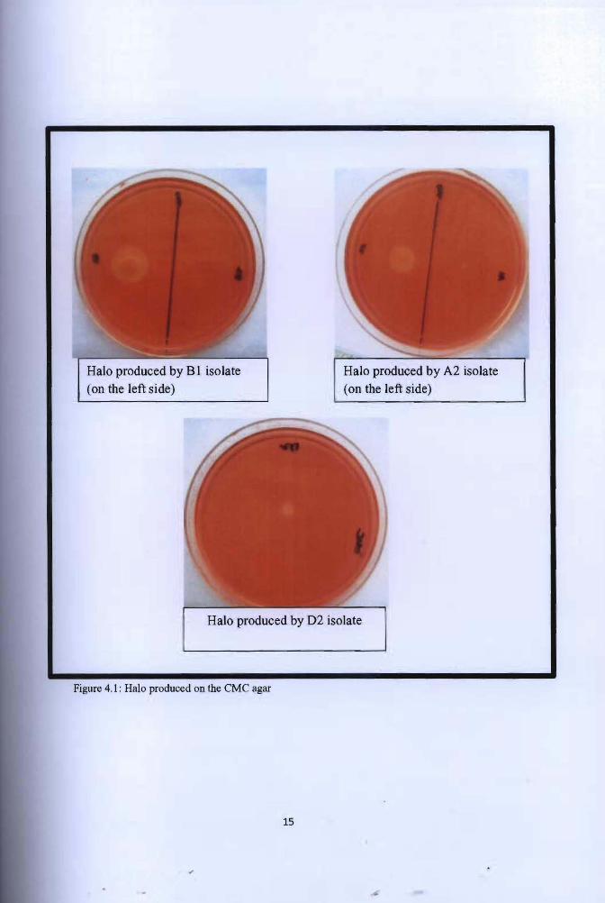
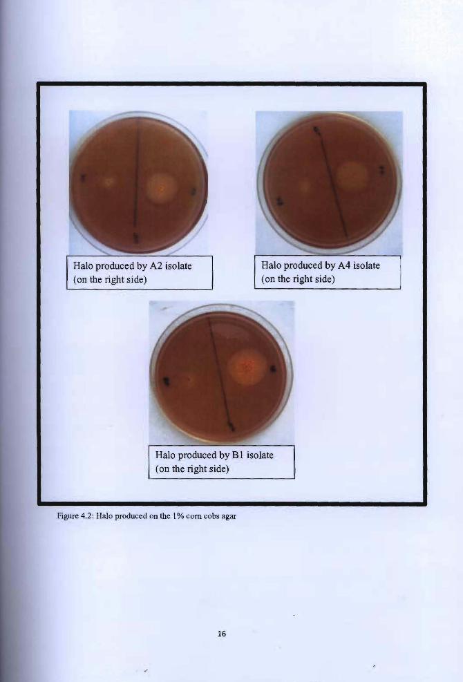
4.2 Secondary screening
Each of the 16 colonies was picked and streaked onto the CMC agar plates and corn cobs agar
plates. The plates were incubated for 24 hours at 37°C. After that, the colonies were flooded
with Congo red solution. Sodium chloride (NaCl) solution was not used to wash the Congo
red staining because the observation was clear enough. Congo red staining procedure stained
the media inside the plates. If the colony produces enzymes, the dye will not stain the media,
thus producing halos, that is the clearing zone around the colony. On the corn cobs plates,
there were three colonies able to produce halo (A2, A4, Bl) while on the CMC plates, there
were also three colonies able to produce halo (A2, B I, D2) (Figure 4 .1 and 4.2). The diameter
of the colony and the halo produced was measured. Table 4.2 and table 4.3 is the measurement
of the halo and its ratio.
14
· I, f ;• . '
--_.... Halo produced by B1 isolate Halo produced by A2 isolate
(on the left side) (on the left side)
Halo produced by D2 isolate
Figure 4.1: Halo produced on the CMC agar
15
t • .'
Halo produced by A2 isolate
(on the right side)
Halo produced by A4 isolate
(on the right side)
Halo produced by Bl isolate
(on the right side)
Figure 4.2: Halo produced on the 1 % com cobs agar
16
I •
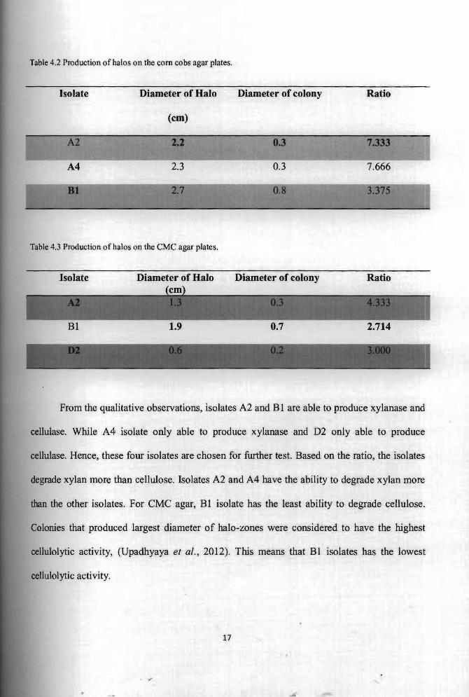
Table 4.2 Production of halos on the com cobs agar plates.
Isolate Diameter of Halo Diameter of colony Ratio
(cm)
A2 0.3
A4 2.3 0.3
Table 4.3 Production of halos on the CMC agar plates.
Isolate Diameter of Halo Diameter of colony Ratio
From the qualitative observations, isolates A2 and Blare able to produce xylanase and
cellulase. While A4 isolate only able to produce xylanase and D2 only able to produce
cellulase. Hence, these four isolates are chosen for further test. Based on the ratio, the isolates
degrade xylan more than cellulose. Isolates A2 and A4 have the ability to degrade xylan more
than the other isolates. For CMC agar, Bl isolate has the least ability to degrade cellulose.
Colonies that produced largest diameter of halo-zones were considered to have the highest
cellulolytic activity, (Upadhyaya et al., 2012). This means that Bl isolates has the lowest
cellulolytic activity.
17
t' i;. . '
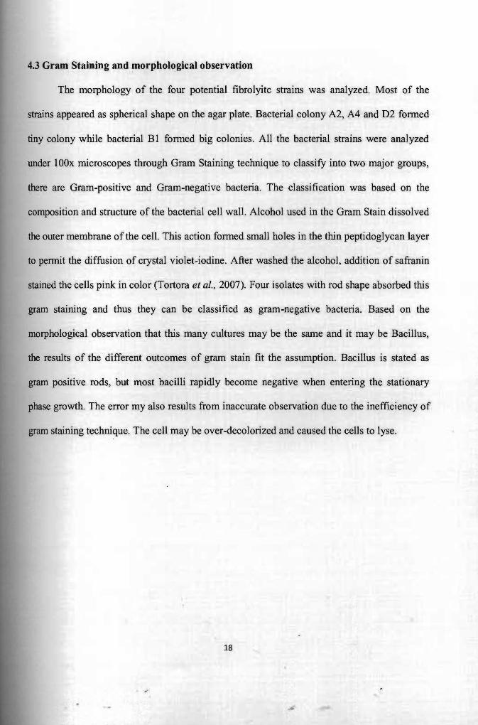
4.3 Gram Staining and morphological observation
The morphology of the four potential fibrolyitc strains was analyzed. Most of the
strains appeared as spherical shape on the agar plate. Bacterial colony A2, A4 and D2 fonned
tiny colony while bacterial BI fonned big colonies. All the bacterial strains were analyzed
under 100x microscopes through Gram Staining technique to classify into two major groups,
there are Gram-positive and Gram-negative bacteria. The classification was based on the
composition and structure of the bacterial cell wall. Alcohol used in the Gram Stain dissolved
the outer membrane of the cell. This action fonned small holes in the thin peptidoglycan layer
to pennit the diffusion of crystal violet-iodine. After washed the alcohol, addition of safranin
stained the cells pink in color (Tortora et ai., 2007). Four isolates with rod shape absorbed this
gram staining and thus they can be classified as gram-negative bacteria. Based on the
morphological observation that this many cultures may be the same and it may be Bacillus,
the results of the different outcomes of gram stain fit the assumption. Bacillus is stated as
gram positive rods, but most bacilli rapidly become negative when entering the stationary
phase growth. The error my also results from inaccurate observation due to the inefficiency of
gram staining technique. The cell may be over-decolorized and caused the cells to lyse.
18