SURVIVAL OF HUMAN PROSTATE CARCINOMA CELL LINE...
37
SURVIVAL OF HUMAN PROSTATE CARCINOMA CELL LINE (DU145) IN THE PRESENCE OF Ficus deltoidea AND Labisia pumila AQUEOUUS EXTRACTS KOSAR ALI OMER UNIVERSITI TEKNOLOGI MALAYSIA
Transcript of SURVIVAL OF HUMAN PROSTATE CARCINOMA CELL LINE...
SURVIVAL OF HUMAN PROSTATE CARCINOMA CELL LINE (DU145) IN
THE PRESENCE OF Ficus deltoidea AND Labisia pumila AQUEOUUS
EXTRACTS
KOSAR ALI OMER
UNIVERSITI TEKNOLOGI MALAYSIA
SURVIVAL OF HUMAN PROSTATE CARCINOMA CELL LINE (DU145) IN
THE PRESENCE OF Ficus deltoidea AND Labisia pumila AQUEOUUS EXTRACTS
KOSAR ALI OMER
A dissertation submitted in partial fulfillment of the requirement for the award of the
degree of Master of Science (Biotechnology)
Faculty of Biosciences and Bioengineering
Universiti Teknologi Malaysia
JANUARY 2013
iii
To my beloved wife (Shaee), father (Ali), mother (Badeaa) and sisters (Kizhan, Zhwan)
and my uncle (Latif)
iv
ACKNOWLEGEMENTS
First and foremost, Alhamdulillah, thanks to Allah, for giving me the strength,
patience and health to go through all obstacles in order to complete this research. With
His blessing, this project is finally accomplished.
I would like to thanks my main supervisor Dr. Mohammed Arshad Javed for his
believing in me and all his supports and guidance by his courageous advices. I would
also want to express my appreciation and thanks to my Co-supervisor Prof. Dr.
Mohammed Roji Sarmidi for all his help during this project and giving me helpful hand
in every step of the work making everything easy for me. I would like to express my
deep appreciation for both my fiends Moh. Mukrish bin Mohd Hanafi and Muhammad
Fauzi Abdjalil efforts, with out their help I couldn’t finish this project as it is now,
special thanks for all the members of Tissue Culture Engineering Laboratory (TCEL) for
letting me work in their laboratory.
I would like to express my grateful to Kurdistan region government, Ministry of
higher education / HCDP, Ministry of health and Hiwa hospital to give me a chance for
completing my post graduate study.
Last but not least, I’m very thankful for my dear wife for all her supports and
encouragements during the time of study. Special thanks for my father and mother for all
their love and prayers. Thanks to all my friends and relatives who support me, especially
my uncle Latif and Dara, Dr. Taher Arif and Miss DeeDee Baumgraner. Thanks a lot for
everything. May God repay your kindness and help in the future.
v
ABSTRACT
Ficus deltoidea and Labisia pumila are two local plants that have been used in
traditional medicine for along time for enhancement reproductive system and cancer
treatment. In this study cell based assay were used to determine the effect of aqueous
extract of these two plants on the Human Prostate carcinoma (DU145) cell line. MTT
assay, viability test and morphological studies were carried out. Ficus deltoidea showed
higher growth inhibitive than Labisia pumila extract. The MTT assay indicated that the
most inhibitive concentration of the extracts is 1*10-3 µg/ml, this concentration is
consistent through out the course of this study. MTT assay demonstrated that Ficus
deltoidea extracts killed around 60% and Labisia pumila around 42% of the cells
compared with the negative control. In the viability test Ficus deltoida also recorded a
better effect than Labisia pumila which killed all the cells with in 7 days. In the case of
Labisia pumila extract is took 9 days, while 12 days needed for negative control. Both
extract showed almost similar effect on the morphology of the cells. Observation under
inverted microscope for both extracts observed irregular detachment and clumping of the
cells. There was morphological changing of the cells from hexagonal to round shape. In
apoptotic body study the effect was similar in blabbing cells, chromatin condensation,
apoptotic cells and late apoptotic body formation, the difference was in the living cells
which for Labisia pumila extract more living cells seen than Ficus deltoidea extract. In
general both aqueous extracts showed a growth suppressive effect on the DU145 cell
line, but Ficus deltoidea extract was more effective than Labisia pumila extract.
vi
ABSTRAK
Ficus deltoidea dan Labisia pumila ialah dua tumbuhan tempatan yang telah
lama digunakan dalam perubatan tradisional untuk meningkatkan sistem pembiakan dan
mengubati penyakit kanser. Penilaian berdasarkan kajian sel ini telah digunakan untuk
menentukan kesan pengekstrakan akueus dari kedua-dua tumbuhan ini ke atas Human
Prostate carcinoma (DU145) garis sel. Sehubungan itu, penilaian MTT, ujian daya maju
dan kajian morfologi telah dijalankan. Ekstrak Ficus deltoidea menunjukkan
peningkatan pembantutan yang lebih tinggi berbanding dengan ekstrak Labisia pumila.
Penilaian MTT menunjukkan bahawa kepekatan pembantutan yang paling tinggi ialah
1*10-3 µg/ml; kepekatan ini sejajar sepanjang kajian ini. Penilaian MTT menunjukkan
bahawa ekstrak Ficus deltoidea menghapuskan lebih kurang 60% daripada sel-sel dan
Labisia pumila lebih kurang 42%; berbanding dengan kawalan negative. Dalam ujian
daya maju, Ficus deltoida juga mencatatkan kesan yang lebih baik berbanding dengan
Labisia pumila, iaitu menhapuskan kesemua sel dalam jangkamasa 7 hari. Ekstrak
Labisia pumila pula mengambil masa 9 hari, sedangkan 12 hari telah diperlukan untuk
kawalan negatif. Kedua-dua ekstrak ini menunjukkan kesan yang hampir sama ke atas
morfologi sel. Penelitian di bawah mikroskop keatas kedua-dua ekstrak ini menunjukkan
pemisahan dan pengelompokan sel-sel yang tidak tetap.Terdapat juga perubahan
morfologi sel-sel tersebut dari bentuk heksagon ke bentuk bulat. Didalam kajian
apoptotik, kesan yang sama ditunjukkan dalam ‘blabbing cells’, kondensasi kromatin,
sel-sel apoptotik dan dalam pembentukan lewat badan apoptik; perbezaannya ialah sel-
sel hidup, iaitu Labisia pumila mengekstrakkan lebih banyak sel-sel hidup berbanding
dengan ekstrak Ficus deltoidea. Secara amnya, kedua-dua ekstrak akueas ini
menunjukkan kesan peningkatan dalam pembantutan keatas DU145 garis sel, tetapi
ekstrak Ficus deltoidea lebih berkesan daripada ekstrak Labisia pumila.
vii
TABLE OF CONTENTS
CHAPTER TITLE PAGE
TITLE i
DECLARATION ii
DEDICATION iii
ACKNOLEDGEMENTS iv
ABSTRACT v
ABASTRAK vi
TABLE OF CONTENTS vii
LIST OF TABLES xiv
LIST OF FIGURES xv
LEST OF SYMBOLS / ABBREVIATIONS xviii
LIST OF APPENDICES xix
1 INTRODUCTION
1.1 Research background 1
1.2 Objective of the study 3
viii
1.3 Scopes of the study 4
1.4 Research contributions 4
2 LITERATURE REVIEW
2.1 Over view 5
2.2 Introduction 5
2.3 Use of Herbs in medicine 6
2.4 Ficus spp. (Fig) and its use in medicine 9
2.4.1 Ficus deltoidea plant and its use in medicine 13
2.5 Labisia pumila 17
2.5.1 Traditional use of Labisia pumila 18
2.5.2 Phytochemical compounds in Labisia pumila 19
2.6 Cancer 20
2.7 Prostate cancer 23
2.7.1 Prostate 23
2.7.2 Prostate cancer 24
2.7.2.1 Prostate cancer symptoms 25
2.7.2.2 Risk factors that enhance Prostate cancer 26
2.7.2.3 Prostate cancer diagnosis 27
ix
2.7.2.3.1 Prostate Specific Antigen 27
2.7.2.3.2 Digital rectal examination 28
2.7.2.3.3 Trans-rectal ultrasound 29
2.7.2.3.4 Needle biopsy 29
2.7.2.4 Prostate cancer grading systems 30
2.7.2.4.1 The Gleason Grading
System
30
2.7.2.4.2 H staging system 31
2.8 Plant and phytochemical compounds against cancer 33
2.9 Cell culture 37
2.9.1 Cell growth 38
2.9.2 Cell death; apoptosis and necrosis 38
2.9.2.1 Determining the cell death 40
2.9.2.1.1 Viability test 41
2.9.2.1.2 The MTT assay 43
2.9.2.1.3 Ethidium bromide and
Acridin orange (EB/AO) double staining
method
45
x
3 METHODOLOGY
3.1 Introduction 48
3.2 Materials 49
3.2.1 Chemical requirements 49
3.2.2 Instruments 49
3.2.3 Cell line 50
3.3 Preparation of extract 51
3.3.1 Preparation of aqueous extracts from F.
deltoidea plant
51
3.3.2 Preparation of aqueous extract from Labisia
pumila plant
51
3.4 Cell culture protocol 52
3.4.1 Cells seeding 52
3.4.2 Medium renewal 52
3.4.3 Cell subculture 53
3.4.4 Cell Cryopreservation 54
3.4.5 Cell Recovery 54
3.4.6 Cell Counting 55
3.4.7 Growth Curve Profile 56
3.5 Cell Proliferation Analysis 57
xi
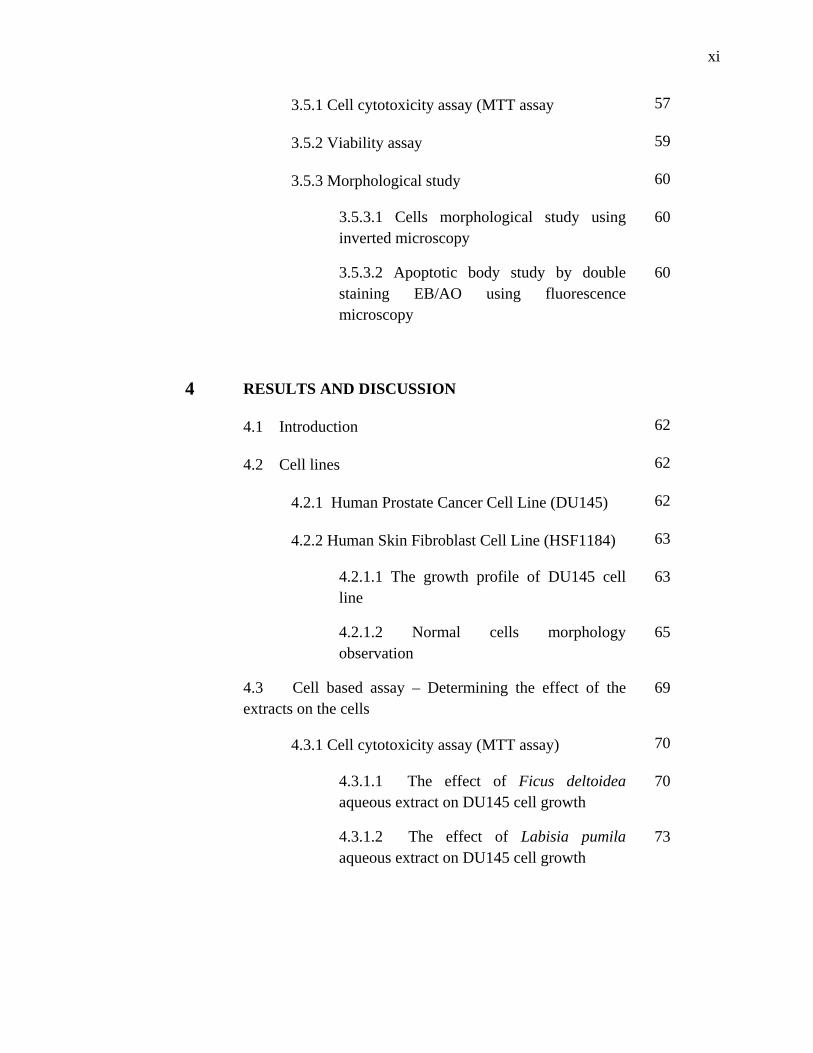
3.5.1 Cell cytotoxicity assay (MTT assay 57
3.5.2 Viability assay 59
3.5.3 Morphological study 60
3.5.3.1 Cells morphological study using inverted microscopy
60
3.5.3.2 Apoptotic body study by double staining EB/AO using fluorescence microscopy
60
4 RESULTS AND DISCUSSION
4.1 Introduction 62
4.2 Cell lines 62
4.2.1 Human Prostate Cancer Cell Line (DU145) 62
4.2.2 Human Skin Fibroblast Cell Line (HSF1184) 63
4.2.1.1 The growth profile of DU145 cell line
63
4.2.1.2 Normal cells morphology observation
65
4.3 Cell based assay – Determining the effect of the extracts on the cells
69
4.3.1 Cell cytotoxicity assay (MTT assay) 70
4.3.1.1 The effect of Ficus deltoidea aqueous extract on DU145 cell growth
70
4.3.1.2 The effect of Labisia pumila aqueous extract on DU145 cell growth
73
xii
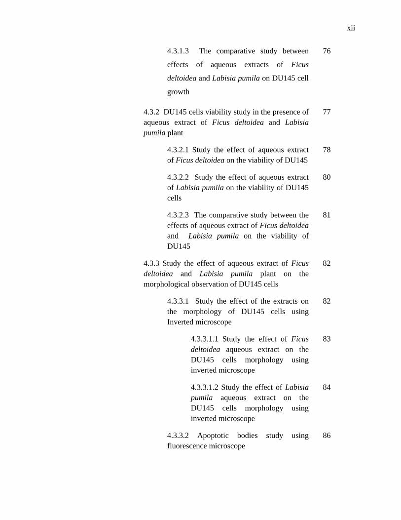
4.3.1.3 The comparative study between
effects of aqueous extracts of Ficus
deltoidea and Labisia pumila on DU145 cell
growth
76
4.3.2 DU145 cells viability study in the presence of aqueous extract of Ficus deltoidea and Labisia pumila plant
77
4.3.2.1 Study the effect of aqueous extract of Ficus deltoidea on the viability of DU145
78
4.3.2.2 Study the effect of aqueous extract of Labisia pumila on the viability of DU145 cells
80
4.3.2.3 The comparative study between the effects of aqueous extract of Ficus deltoidea and Labisia pumila on the viability of DU145
81
4.3.3 Study the effect of aqueous extract of Ficus deltoidea and Labisia pumila plant on the morphological observation of DU145 cells
82
4.3.3.1 Study the effect of the extracts on the morphology of DU145 cells using Inverted microscope
82
4.3.3.1.1 Study the effect of Ficus deltoidea aqueous extract on the DU145 cells morphology using inverted microscope
83
4.3.3.1.2 Study the effect of Labisia pumila aqueous extract on the DU145 cells morphology using inverted microscope
84
4.3.3.2 Apoptotic bodies study using fluorescence microscope
86
xiii
5 CONCLUSIONS AND RECOMENDATION
5.1 The conclusion of this study 89
5.2 Recommendations 91
REFERENCES 92
APPENDICES 110
xiv
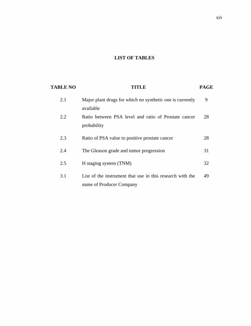
LIST OF TABLES
TABLE NO TITLE PAGE
2.1 Major plant drugs for which no synthetic one is currently
available
9
2.2 Ratio between PSA level and ratio of Prostate cancer
probability
28
2.3 Ratio of PSA value to positive prostate cancer 28
2.4 The Gleason grade and tumor progression 31
2.5 H staging system (TNM) 32
3.1 List of the instrument that use in this research with the
name of Producer Company
49
xv
LIST OF FIGURES
FIGURE NO TITLE PAGE
2.1 Male “mas cotek” leave 14
2.2 Female “mas cotek” leave 14
2.3 Labisia pumila var. alata 17
2.4 Cancer Hallmarks 21
2.5 Prostate gland and surrounding organs 23
2.6 Statistics for prostate cancer in different countries and
the rate of mortality among them
25
2.7 The steps and mechanisms of necrosis and apoptosis 40
2.8 Cell growth curve in cell culture 42
2.9 Reaction happen on MTT solution to purple formazan by
mitochondrial reductase enzyme
44
3.1 The design of overall experimental procedure 47
3.2 Hemocytometer, magnified view of one square that
shows the cells that can be count and those that
neglected. The clear rings are living cells and the dark
rings are dead cells
55
xvi
4.1 The complete growth profile of DU145 cell line 64
4.2 The morphology of DU145 cell line on (a) day1, (b) day
3, (c) day 6, (d) day 8 and (e) day 14 using 10X
magnification power of inverted microscope
69
4.3 Effect of aqueous extract of Ficus deltoidea plant at
different concentration on DU145 cell
70
4.4 Comparative study of the effect of aqueous extract of
Ficus deltoidea plant at different concentration on
HSF1184 and DU145 cells
71
4.5 Effect of aqueous extract of Labisia pumila plant at
different concentration on DU145 cell
72
4.6 Comparative study of the effect of aqueous extract of
Labisia pumila plant at different concentration on
growth of HSF1184 and DU145 cells
74
4.7 Comparative study of the effect of aqueous extract of
Ficus deltoidea and Labisia pumila plant at different
concentration on growth of DU145 cells
76
4.8 The effect of Ficus deltoidea aqueous extract on the
viability of DU145 cell compared with the normal
viability of the cells
78
4.9 The effect of Labisia pumila aqueous extract on the
viability of DU145 cell compared with the normal
viability of the cells
80
4.10 Comparative study between the effects of aqueous
extract of Ficus deltoidea and Labisia pumila on the
viability of DU145 cells
81
xvii
4.11 Changes in cells morphology in different times of extract
effect (a) 1st day of exposure, (b) 4th day of the extract
exposure and (c) 7th day of exposure under inverted
microscope with 10X power
83
4.12 Changes in cells morphology in different times of extract
effect (a) 1st day of exposure, (b) 4th day of the extract
exposure and (c) 9th day of exposure under inverted
microscope with 10X power
85
4.13 The morphology of DU145 cell stained with EB/AO and
observed under the fluorescence microscope after (a)
cells with out extract (control), (b) cells after treatment
with 0.001 µg/ml Ficus deltoidea aqueous extract,
(c) cells after treatment with 0.001µg/ml Labisia pumila
aqueous extract
87
xviii
LIST OF ABBREVIATIONS
ATCC - American Type Culture Collection
AO - Acridine orange
CO2 - Carbon dioxide
DMEM - Dulbeco Minimum Essential Medium
DMSO - Dimethyle Sulfoxide
EB - Ethedium bromid
ECACC - European Collection of Cell Cultures
EDTA - Ethylene Diamine Tetra Acidicacid
FBS - Foetal Bovine Serum
HSF - Human Skin Fibroblast
MARDI - Malaysian Agricultural Research and
Development Institute
MTT - 3-(4,5 Dimethylthiozol-2-yl)-2,5-
diphenyletetrazolium
PBS - Phosphate Buffer Saline
PDT - Population Doubling Time
xix
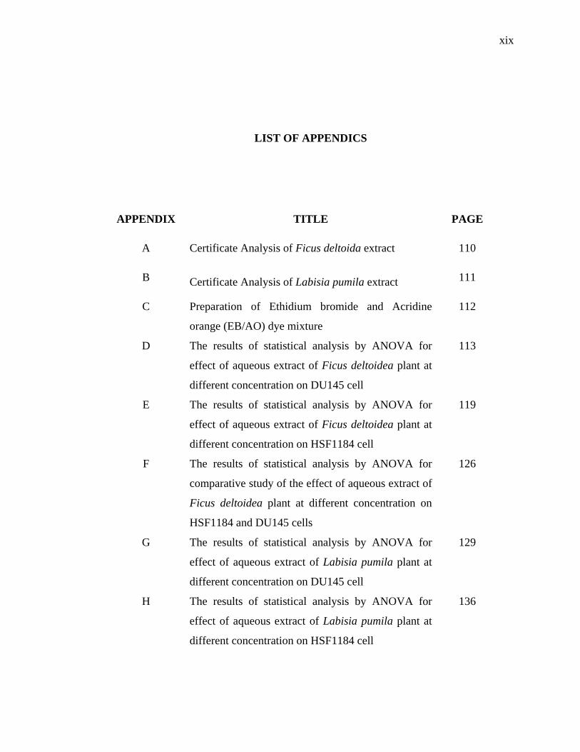
LIST OF APPENDICS
APPENDIX TITLE PAGE
A Certificate Analysis of Ficus deltoida extract 110
B Certificate Analysis of Labisia pumila extract 111
C Preparation of Ethidium bromide and Acridine
orange (EB/AO) dye mixture
112
D The results of statistical analysis by ANOVA for
effect of aqueous extract of Ficus deltoidea plant at
different concentration on DU145 cell
113
E The results of statistical analysis by ANOVA for
effect of aqueous extract of Ficus deltoidea plant at
different concentration on HSF1184 cell
119
F The results of statistical analysis by ANOVA for
comparative study of the effect of aqueous extract of
Ficus deltoidea plant at different concentration on
HSF1184 and DU145 cells
126
G The results of statistical analysis by ANOVA for
effect of aqueous extract of Labisia pumila plant at
different concentration on DU145 cell
129
H The results of statistical analysis by ANOVA for
effect of aqueous extract of Labisia pumila plant at
different concentration on HSF1184 cell
136
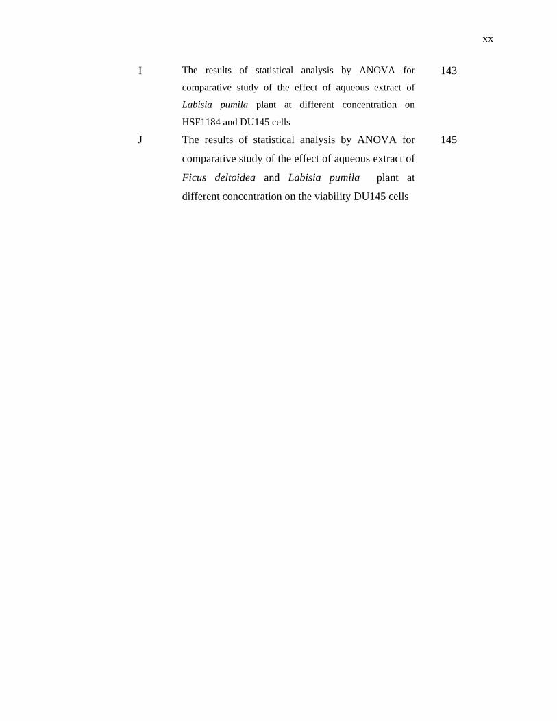
xx
I The results of statistical analysis by ANOVA for
comparative study of the effect of aqueous extract of
Labisia pumila plant at different concentration on
HSF1184 and DU145 cells
143
J The results of statistical analysis by ANOVA for
comparative study of the effect of aqueous extract of
Ficus deltoidea and Labisia pumila plant at
different concentration on the viability DU145 cells
145
CHAPTER 1
INTRODUCTION
1.1 Research background
Prostate cancer is one of the common diagnosed cancer worldwide, especially
in western societies. In the United States, there were 32,050 deaths recorded out of
217,730 newly diagnosed cancer in 2010 (American Cancer Society, 2012).
Approximately one in seven men in America will be diagnosed with prostate cancer
and this makes it as the most common cancer in the United States.
In Europe, 3.2 million new cases of cancer are diagnosed and 1.7 million
deaths due to cancer recorded in 2008. Out of this, Prostate cancer comprised around
11.9 % of all male cancer and around 6 % of all cancer deaths among men caused by
prostate cancer in the European Union (Ferlay et al., 2010).
Prostate cancer is the fourth common cancer in Malaysia among men. There
were 502 prostate cancer cases diagnosed in 2007 and reported to NCR. The
incidence of prostate cancer increases after the age of 45 years and higher in Chinese
male compared to Malay and Indian. Of the cases with reported stage, 40.6% of the
cases were diagnosed at stage I and II (Zainal and Nor Saleha, 2011).
2
According to GLOBOCAN 2008; (a project of the International Agency for
Research on Cancer), Prostate cancer was the second most diagnosed cancer among
male, the 5th common cancer overall, and the 6th cause of death in men from cancer
worldwide. Recently, GLOBOCAN announced that around 258,000 patients died
due to prostate cancer in 2008, with an estimated 899 000 new cases (Ferlay et al.,
2010).
Novel anticancer agents are needed because many cancer patients developed
resistance to standard anticancer agents during treatment. In addition, these agents
are toxic causing undesired side effect and also hypersensitive reactions. Presently,
less than 1% of the estimated 250000 higher plant species on earth has been
investigated chemically for their bioactivity. There are still possibilities of finding
novel compounds with desired bioactivity (Nor Azurah, 2011).
Several species of Fig tree showed to have multiple cancer preventive, anti-
inflammatory and cancer therapeutic effects. Ficus deltoidea is one of these species
of Fig trees “or as locally known as Mas Cotek” from the family Moraceae. Recently
this herb attracts many researchers who are interested in traditional medicine and
phytotherapy. Its therapeutic effect includes regulating blood circulation, and also
traditionally its leaf is used by female as post partum medication. Compared to other
traditional Malaysian herbs, this plant species receives little scientific research and
the information regarding is very limited.
Studies have shown that Labisia pumila plant extract contains certain
phytochemicals which have great potential in cancer prevention specifically prostate
cancer. Flavonoids and other phenolic acids are compounds that are found in the
plant extract in remarkable amount are responsible for the wide spectrum of
pharmacological activities attributed to the herb (Zhang et al., 2008). Even though,
no specific research has been done to investigate the anticancer properties of Labisia
pumila extract, the presence of these phytochemicals could lead to the finding of
another great potential of the plant. Other researches using plant extract with similar
phytochemical compounds have clearly displayed their anticancer properties by
3
addressing some of the hallmarks of cancer. Gallic acid is identified as the major
anticancer compound in T.sinesis leaf extract. Studies done using this extract showed
that gallic acid is cytotoxic to DU145 prostate cancer cells through generation of
reactive oxygen species (ROS) (Huei et al., 2009).
Cell based assay is increasingly used in the recent years for determination of
bioactivity of plant extracts. Simplicity of homogenous methods and correlation with
in vitro cytotoxicity data made this assay as an alternative method for animal testing
in toxicology laboratories. Cell culture technology enables us to investigate the effect
of plant extract on cell viability or cytotoxicity in vitro and also observation of cell
morphology (Freshney, 2005).
There are several ways to determine cell viability and observation of cell
morphology. Staining and observation of cell morphology under light or fluorescence
microscope is one of these methods and molecular method is another one by using
DNA fragmentation screening we can distinguish between the cells. MTT assay is a
standard colorimetric method that can be used for determining the biochemical
activity of the extracts by distinguishing between the live and dead cells and when
there was any disruption of critical biochemical function (Lobnar, 2000).
1.2 Objective of study
The objective of this study is to establish the effect of aqueous extract of
Ficus deltoidea and Labisia pumila on the growth and survival of prostate carcinoma
cell line (DU145).
4
1.3 Scopes of the study
The scopes of this study are:
1. To investigate the effect of Ficus deltoidea aqueous extracts on the
characteristics and the morphology of human prostate carcinoma cell
line.
2. To investigate the effect of Labisia pumila aqueous extracts on the
characteristics and the morphology of human prostate carcinoma cell
line.
3. To compare the effect of Ficus deltoedia aqueous extract with the
effect of Labisia pumila aqueous extract on the Human Prostate
carcinoma cell line.
1.4 Research contributions
This research contributed in the finding the importance of Ficus deltoidea and
Labisia pumila extracts as a medicinal and anticancer agents against prostate cancer.
This study maybe counted as the basic steps in finding novel drugs for prostate
cancer cell, which will pave a way for future studies especially in mechanism of
action of these extracts and isolating the active phytochemical compounds in the
extracts in order to treat this disease at its early stages.
92
REFERENCES
Abad-Garcia B., Berrueta L.A., Garmon-Lobato S., Gallo B. and Vicente F. (2009).
general analytical strategy for the characterization of phenolic compounds in
fruit juices by high-performnace liquid chromatography with diode array
detection coupled to electrospray ionization and triple quadrupole mass
spectrometry. J. Chromatol. 1216, 5398–5415.
Abdullah NA., Karsani SA. and Aminudin N. (2008). Effects of Ficus deltoidea extract
on the serum protein profile of simultaneously hypertensive rats (SHR). J
Proteomics and Bioinformatics. S2, 143.
Abukakar MG., Ukwuani AN. and Shehu RA. (2008). Phytochemical screening and
antibacterial activity of Tamarindus indica Pulp Extract. J. Biochem. 3(2), 134-
138.
Ackernecht E.H. (1973). Therapeutics: From the Primitives to the Twentieth Century.
New York: Hafner Press.
Adlercreutz H. (2002). Phyto-oestrogens and cancer. Lancet Oncol. 364-373.
Adrian G., Rachel B., Charles Y. (1999). Induction of apoptosis in prostate cancer cell
lines by the green tea component, (-)-epigallocatechin-3-gallate. Cancer Letters.
130, 1–7.
Advisory Committee on Cancer Prevention (2000). Recommendations on cancer
screening in the European Union. Eur J Cancer. 36, 1473–8.
Agrawal S., Agarwal SS. (1990). Preliminary observations on leukaemia specific
agglutinins from seeds. Indian Journal of Medical Research 92, 38–42.
Ahmad I., Aqil F. and Owais M. (2006). Modern Phytomedicine. Turning Medicinal
Plants into Drugs. India: Wiley-VCH.
Ahmedin J., Freddie B., Melissa M., Jacques F., Elizabeth W., David F. (2011). Global
Cancer Statistics. CA - A Cancer Journal for Clinicians. 61, 69–90.
93
Aiyegoro O., Okoh A. (2009). Use of bioactive plant products in combination with
standard antibiotics: implications in antimicrobial chemotherapy. Journal of
Medicinal Plants Research 3, 1147–1152.
Aiyer K. N. and Kolammal M. (1960). Pharmacognosy of Ayurvedic drugs. Trivandrum,
India: University of Kerala.
Ali Z. and Khan IA. (2011). Alkyl phenols and saponins from the roots of Labisia
pumila (Kacip Fatimah). Phytochem. 72, 2075–80.
Allen EA, Kahane H and Epstein JI. (1998). Repeat biopsy strategies for men with
atypical diagnoses on initial prostate needle biopsy. Urology. 52:803-807.
American Cancer Society (2012). Cancer Facts & Figures. Atlanta, Ga: American
Cancer Society.
American Society of Clinical Oncology (2011). The history of Cancer. Retrieved on
October 11, 2012. from www.bordet.be/en/presentation/history/cancer_e/ cancer1
.htm.
Amin A., Nanji M.D. and Susanne H. (1997). Apoptosis and necrosis two types of cell
death in alcoholic Liver disease. Alcohol and research world. 21, 4-10.
Anon (2006). Mas Cotek kini herba terpilih. D’herba, 4, 11.
Atawodi S.E., Ameh D.A., Ibrahim S., Andrew J.N., Nzelibe H.C., Onyike E.O., Anigo
K. M., Abu E.A., James D.B., Njoku G.C. and Sallau A.B. (2001). Indigenous
Knowledge System for Treatment of Trypanosomiasis in Kaduna State of
Nigeria. J. Ethnopharmacol. 79, 279-282.
Aus G., Ahlgren G., Hugosson J., Pedersen KV., Rensfeldt K. and Soderberg R. (1997).
Diagnosis of prostate cancer: optimal number of prostate biopsies related to
serum prostate-specific antigen and findings on digital rectal examination. Scand
J Urol Nephrol. 31(6), 541-544.
Aus G., Becker C., Franzén S., Lilja H., Lodding P. and Hugosson J. (2004). Cumulative
prostate cancer risk assessment with the aid of the free-to-total prostate specific
antigen ratio. Eur Urol. 45(2), 160-165.
Avula B., Wang YH., Ali Z., Smillie TJ. and Khan IA. (2011). Quantitative
determination of triterpene saponins and alkenated-phenolics from Labisia
pumila using an LC–UV/ELSD method and confirmation by LC–ESI-TOF.
Planta Med. 77, 1742–8.
94
Baker J. T., Borris R. P., Carte B., Cordell G. A., Soejarto D., Cragg G. M., Gupta M. P.,
Iwu M. M., Madulid D. R. and Tyler V. E. (1995). Natural product drug
discovery and development: new perspectives on international collaboration. J.
Nat. Prod. 58, 1325-1357.
Balandrin M. F., Kinghorn A. D. and Farnsworth N. R. (1993). Chapter 1. In Kinghorn
A. D., Balandrin M. F., (Eds.). Human Medicinal Agents from Plants (p. 2-12).
Washington: American Chemical Society.
Bentley R. (1990). The shikimate pathway—a metabolic tree with many branches.
Critical Reviews in Biochemistry and Molecular Biology. 25, 307–384.
Berg CC. (1989). Classification and distribution of Ficus. Experientia. 45, 605-611.
Black D. (1997). One Hundred Years of Aspirin. The Lancet. 350(9075), 437–439.
Black RJ., Bray F., Ferlay J. and Parkin DM. (1997). Cancer incidence and mortality in
the European Union: cancer registry data and estimates of national incidence for
1990. Eur J Cancer. 33(7), 1075-1107.
Bowman T. and Zon L. (2010). Swimming into the future of drug discovery: in vivo
chemical screens in zebrafish. ACS Chem. Biol. 5, 159–161.
Brahmachari H., Augusti K. (1962). Orally effective hypoglycemic agents from plants.
Journal of Pharmacy and Pharmacology. 14, 254–255.
Bray F., Sankila R, Ferlay J. and Parkin DM. (2002). Estimates of cancer incidence and
mortality in Europe in 1995. Eur J Cancer. 38(1), 99-166.
Brickell C. and J. D. Zuk (1997). The American Horticultural Society A-Z Encyclopedia
of Garden Plants. New York: DK Publishing, Inc.
Buhler D. and Cristobal, M. (2000). Antioxidant activities of flavonoids. Retrieved on
October 11, 2012. From http://lpi.oregonstate.edu/f-w00/flavonoid.html.
Burant C., Takeda J., Brot-Laroche E., Bell G. and Davidson N. (1992). Fructose
transporter in human spermatozoa and small intestine is GLUT5. J. Biol. Chem.
267, 14523-14526.
Burkill H.M. (1997). The useful plants of West Tropical Africa. (2nd Edition). Volume 4,
Families M–R. Royal Botanic Gardens (pp. 969). United Kingdom: Kew
publishing.
Burkill IH. (1966). A dictionary of the economic product of the Malay Peninsula. Vol.II
(I–Z). Kuala Lumpur: Government of Malaysia and Singapore by the Ministry of
Agriculture and Cooperative.
95
Bustamanate E., and Pedersen P. (1977). High aerobic glycolysis of rat hepatoma cells in
culture: Role of mitochondrial hexokinase. Proc. Natl. Acad. Sci. USA. 74 (9),
3735-3739.
Butler M. (Ed.) (2004). Animal cell culture and technology. London and New York:
Graland science/BIOS scientific publisher.
Carvalhal GF., Smith DS., Mager DE. and Catalona WJ. (1999). Digital rectal
examination for detecting prostate cancer at prostate specific antigen levels of 4
ng/mL or less. J Urol. 161(3), 835-839.
Catalona W. (1996). Prostate cancer screening. BJU Int. 94(7), 964-6.
Changwei A., Anping Li, Abdelnaser A. Elzaawely, Tran D. Xuan, Shinkichi Tawata
(2007). Evaluation of antioxidant and antibacterial activities of Ficus microcarpa
L. fil. Extract. Food Control. 19, 940–948.
Chiang, YM., Chang, JY., Kuo CC., Chang CY., Kuo YH. (2005). Cytotoxic triterpenes
from the aerial roots of Ficus microcarpa. Phytochemistry. 66, 495–501.
Choi H.K., Kim D.H., Kim J.W., Ngadiran S., Sarmidi M.R. and Park C.S. (2010).
Labisia pumila extract protects skin cells from photoaging caused by UVB
irradiation. J. Biosci. Bioeng. 109, 291–296.
Christofk HR, Vander Heiden MG, Wu N, Asara JM, Cantley LC (2008). Pyruvate
kinase M2 is a phosphotyrosine-binding protein. Nature. 452, 181-186.
Chung LW., Baseman A., Assikis V. and Zhau HE. (2005). Molecular insights into
prostate cancer progression: the missing link of tumor microenvironment. J Urol.
173(1), 10-20.
Clark A. M. (1996). Natural products as resources for new drugs. Pharm. Res.13(8),
1133-44.
Cohen J. (1993) Apoptosis. Immunol Today. 14,126-130.
Coligan JE, Kruisbeek AM., Margulis DH, Shevach EM. and Strober W. (1995). Current
Protocols in Immunology. New York, NY: John Wiley.
Cragg G. M., Boyd M. R., Cardellina II J. H., Grever M. R., Schepartz S. A., Snader K.
M. and Suffness M. (1993). Chapter 7. In Kinghorn A.D., Balandrin M. F.
(Eds.). Human Medicinal Agents from Plants (p. 80 – 95). Washington:
American Chemical Society.
Cragg G. M., Newman D. J. and Snader K. M. (1997). Natural products in drug
discovery and development. J. Nat. Prod. 60, 52-60.
96
Daniel S.A., Elizabeth A.C., Mario F. and Joshua A.B. (2008). Epigallocatechin-3
gallate (EGCG) inhibits PC-3 prostate cancer cell proliferation via MEK-
independent ERK1/2 activation. Chemico-Biological interactions. 171, 89-95.
Dey A. C. (1980). Indian Medicinal Plants Used in Ayurvedic Preparations. DehraDun,
India: Bishen Singh Mahendra Pal Singh.
Douglas H. and Robert A. (2011). The hallmarks of Cancer: the next generation. Cell.
144.
Esposti PL, Elman A and Norlen H. (1975). Complications of transrectal aspiration
biopsy of the prostate. Scand J Urol Nephrol. 9(3), 208-213.
Faezeh I. (2008). The effect of Ficus deltoidea’s fruit extraction in different
concentration on mouse Oocyte. Bachelor’s degree. Universiti Teknologi
MARA.
Farnsworth N. R., Akerele O., Bingel A., Soejarto D. and Guo Z. (1985). Medicinal
plants in therapy. Bulletin of the World Health Organization. 63, 965-981.
Fedi P., Tronick S.R. and Aaronson S.A. (1997). Growth factors. in J.F. Holland, R.C.
Bast, D.L. Morton, E. Frei, D.W. Kufe, and R.R. Weichselbaum (eds.). Cancer
Medicine (pp. 41–64). Baltimore, US: Williams and Wilkins.
Fellows L. and Scofield A. (1995). Intellectual Property Rights and Biodiversity
Conservation—An Interdisciplinary Analysis of the Values of Medicinal Plants.
In T. Swanson (ed.). Chemical Diversity in Plants (pp. 19–44). New York:
Cambridge University Press.
Ferlay J., Shin HR., Bray F., Forman D., Mathers CD., Parkin D. (2010). Estimates of
worldwide burden of cancer in 2008: GLOBOCAN 2008. International Journal
of Cancer. 12(127). Wiley online liberary.
Finley J.W. (2003). The antioxidant responsive element (ARE) may explain the
protective effects of cruciferous vegetables on cancer. Nutrition Reviews. 61,
250–254.
Fitzpatrick J. (2004 ). PSA screening for prostate cancer. Urol News. 9 (1), 6-9.
Frankfurt O.S. (1994). Detection of apoptotic leukemic and breast cancer cells with
monoclonal antibody to single-stranded DNA. Anticancer Res. 14,1861-1870.
Franky L., Choi H., Chen Z., Peter S. and Huang Y. (2000). Induction of apoptosis in
prostate cancer cell lines by avonoid, baicalin. Cancer Letters. 160, 219-228.
Freshney I. (2005). Culture of animal cells: A manual of basic technique (5th ed.). USA:
John wiley & sons, Inc.
97
Frezza, C. and Gottlieb E. (2009). Mitochondria in cancer: not just innocent by standers .
Semin. Cancer Biol. 19, 4–11.
Gerber GS. and Chodak GW. (1991). Routine screening for cancer of the prostate. J
Natl Cancer Inst. 83(5), 329-335.
Gleason DF. (1992). Histologic grading of prostate cancer: a perspective. Human
Pathology. 23(3):273.
Govil JN., Singh VK. and Shameema H. (1993). Glimpses in Plant Research Vol. X.
Medicinal Plants: New Vistas of Research (part 1) (p. 69). New Dehli, India:
Today & Tomorrow’s Printers and Publishers.
Hadijah H., Normah A., Ahmad Tarmizi S. and Aida M. (2007). Cholesterol lowering
effect of mas cotek tea in hypercholesterolemic rats. The 2nd International
conference of east-west perspective of functional food science. 5–7 Nov. 2007,
Kuala Lumpur.
Hartwell J.L. (1967). A survey for Plants used against cancer. Lloydia. 30, 379–436.
Hatanaka M. (1974). Transport of sugars in tumor cell membranes. Biochim. Biophys.
Acta. 355, 77-104.
Heidenreich A., Aus G., Abbou C., Bolla M., Joniau S., Matveev V., Schmid H. and
Zattoni F. (2007). Guidelines on Prostate Cancer. European Urology. 53, 68-80.
Heyninck K. and Beyaert R. (2001). Crosstalk between NF-kappa B-activating and
apoptosis-inducing proteins of the TNF-receptor complex. Mol Cell Biol Res
Commun. 4, 259-265.
Huei M.C., Yang C.W., Yi C.C, Chang F.R., Hsu H.K., Hsieh Y.C., Chen C.C. and
Yuan S.S. (2009). Gallic acid, a major component of Toonasinensis leaf extracts,
contains a ROS-mediated anti-cancer activity in human prostate cancer cells.
Cancer letter 286, 161-171.
Husain A., Virmani OP., Popli SP., Misra LN., Gupta MM., Srivastava GN., Abraham Z.
and Singh AK. (1992). Dictionary of Indian Medicinal Plants (p. 546). Lucknow,
India: CIMAP.
Hussain M., Banerjee M. and Sarkar F.H. (2003). Soy isoflavones in the treatment of
prostate cancer. Nutr Cancer. 47, 111-117.
Hyla Cass (2004). Herbs for the Nervous System: Ginkgo, Kava, Valerian,
Passionflower. Semin Integr Med Elsevier Inc. 2, 82-88.
98
Ibrahim M.H., Jaafar H.Z.E., Rahmat A. and Zaharah A.R. (2011). The relationship
between phenolics and flavonoid production with total non structural
carbohydrate and photosynthetic rate in Labisia pumila Benth. Under high CO2
and nitrogen fertilization. Molecules. 16, 162–174.
Jamal JA., Houghton PJ., Milligan SR. and Jantan I. (2003). The oestrogenic and
cytotoxic effects of the extracts of Labisia pumila var. alata and Labisia pumila
var. pumila in vitro. Sains Kesihatan.1, 53–60.
Jemal A., Siegel R. and Ward E. (2006). Cancer statistics. CA Cancer J Clin. 56, 106 -
130.
Kacinski B. and Flick M. (2001). Apoptosis and cutaneous T cell lymphoma. Ann NY
Acad Sci. 941, 194-199.
Karimi E., Jaafar HZE. and Ahmad S. (2011). Phytochemical analysis and anti-microbial
activities of methanolic extracts of leaf, stem and root from different varieties of
Labisa pumila Benth. Molecules. 16, 4438–50.
Kessler D.A. (2000). Cancer and herbs. New England Journal of Medicine. 342, 1742–
1743.
Kirby RS., Christmas TJ., and Brawer MK. (1996). Prostate cancer. London: Times
Mirror International Publishers.
Kirtikar K.R. and B.D. Basu (1996). Indian Medicinal Plants. (2nd Ed.) (pp: 61-62).
Allahabad: Indian Press.
Kirtikar KR and Basu BD (1996). Indian Medicinal Plants. Vol. II, Dehradun, India:
International Book Distributers.
Kolonel LN., Altshuler D. and Henderson BE. (2004). The multiethnic cohort study:
exploring genes, lifestyle and cancer risk. Nat Rev Cancer. 7, 519-527.
Kris-Etherton P.M., Hecker K.D., Bonanome A., Coval S.M., Binkoski A.E., Hilpert
K.F., Griel A.E. and Etherton, T.D. (2002). Bioactive compounds in foods: their
role in the prevention of cardiovascular disease and cancer. American Journal of
Medicine. 113 (Suppl. 9B), 71S–88S.
Kumar S., Shukla Y. N., Lavania U. C., Sharma A. and Singh A. K. (1997). Medicinal
and Aromatic Plants: Prospects for India. J. Med. Arom. Pl. Sc. 19 (2), 361-365.
Kuo, Y. H. and Li Y. C. (1997). Constituents of the bark of Ficus microcarpa L.f.
Journal of the Chinese Chemical Society. 44, 321–325.
99
Lee B.W., Lee J.H., Lee S.T., Lee H.S., Lee W.S., Jeong T.S., Park K.H. (2005).
Antioxidant and cytotoxic activities of xanthones from Cudrania tricuspidata.
Bioorganic & Medicinal Chemistry Letters.15, 5548–5552.
Lee K.H. (1993). Chapter 12. In Kinghorn A. D. and Balandrin. M. F. (Eds.). Human
Medicinal Agents from Plants (p. 170-190). Washington DC: American Chemical
Society.
Lee S., Sze Y., Norhanisah A. and Mohamad R. (2012). Review on Labisia pumila
(Kacip Fatimah): Bioactive phytochemicals and skin collagen synthesis
promoting herb. Fetoterapia journal. 10(1016), 04.002.
Li X. and Darzynkiewicz Z. (1995). Labelling DNA strand breaks with BdrUTP.
Detection of apoptosis and cell proliferation. Cell Prolif. 28, 571.
Lobner D. (2000). Comparison of the LDH and MTT assays for quantifying cell death:
validity for neuronal apoptosis?. J. Neurosci. Methods. 96, 147-152.
MARDI Malaysian Agriculture Research and Development Institute. (2007a).
Functional drink from Mas cotek cultivated MFD4 [Borchure]. Kuala Lumpur:
MARDI.
McChesney J. (1996). Medicinal Resources of the Tropical Forest -Biodiversity and its
Importance to Human Health. In M.J. Balick, E. Elisabetsky and S.A. Laird
(eds.). Biological Diversity, Chemical Diversity, and the Search for New
Pharmaceuticals (pp. 11–18). New York: Columbia University Press.
Mettlin C., Murphy GP., Babaian RJ., Chesley A., Kane RA., Littrup PJ., Mostofi FK,
Ray PS., Shanberg A. and Toi A. (1996). The results of a five-year early prostate
cancer detection intervention. Investigators of the American Cancer Society
National Prostate Cancer Detection Project. Cancer. 77(1), 150-159.
Meyer F., Bairati I., Shadmani R., Fradet Y. and Moore L. (1999). Dietary fat and
prostate cancer survival. Cancer Causes Control. 10(4), 245-251.
Migliaccio S. and Anderson J.B. (2003). Isoflavones and skeletal health: Are these
molecules ready for clinical application. Osteoporos. Int. 14, 361–368.
Mohan G.K., E. Pallavi, B.R. Kumar, M. Ramesh and S. Venkatesh (2007). Hepato-
protective activity of Ficus carica Linn. leaf extract against carbon tetrachloride-
induced hepatotoxicity in rats. DARU. 15, 162-166.
Mukrish H. (2011). The photoprotective and collagen stimulatory effects of Labisia
pimila var pumila extract on UVB irradiated Human skin fibroblast (HSF1184).
Master degree. Universiti Teknologi Malaysia.
100
Naghma Khan and Sarwat Sultana (2005). Modulatory Effect of Ficus racemosa:
Diminution of potassium Bromate-Induced Renal Oxidative Injury and Cell
Proliferation Response. Basic Clin Pharmacol Toxicol. 97(5), 282 – 288.
National Cancer Institute (2008). What you want to know about prostate cancer.
Retrieved on July 4, 2012. US department of health and Human services,
Available at http://www.cancer.gov/espanol.
Nieweg O., Kim E., Wong W., Broussard W., Singletary S., Hortbagyi G., Tubury R.
(1993). Positron emission tomography with uorine-18-deoxyglucose in the
detection and staging of breast cancer. Cancer. 71, 3920-3923.
Nor Azura M. A. (2011). The survival and growth of human ovarian carcinoma cell line
post treatment with Ficus deltoidea extracts. Master degree. Universiti
Teknologi Malaysia.
Nor Azurah M., Lee S., Fadzilah A., Mohamad Roji S. (2011). Cytotoxicity of Aqueous
and Ethanolic Extracts of Ficus deltoidea on Human Ovarian Carcinoma Cell
Line. British Journal of Medicine & Medical Research. 1(4), 397-409.
Pandey G. and Madhuri S. (2009). Some medicinal plants as natural anticancer agents
Pharmacogn Rev. 3, 259–63.
Parekh J. and Chanda SV. (2006). In vitro antimicrobial activity and phytochemical
analysis of some Indian medicinal plants. Indian J. Pharmacol. 31, 53-58.
Park D. J., and Patek P. Q. (1998). Detergent and enzyme treatment of apoptotic cells for
the observation of DNA fragmentation. Biotechniques. 24, 558-560.
Parkin DM., Bray FI. and Devesa SS. (2001). Cancer burden in the year 2000: the global
picture. Eur J Cancer. 37(8), S4-66.
Patrick EM., Armen M. and Gretchen RH (2009). Study: Herbs added to 5,100-year old
Egyptian wine Ancient. Egyptian herbal wines, retrieved on October 15, 2012.
PNAS website. LD News website.
Paula S. (2009). Men and Prostate gland. Retrieved on November 13, 2012 from
www.kiwifamilies.co.nz/articles/men-and-prostrate-glands.
Pezzuto, J.H. (1997). Plant-derived anticancer agents. Biochemical Pharmacology. 53,
121-133.
Plumb J. A. (1999). Cell sensitivity assays, the MTT assay. In brown R. and Boger
Brown U. (Eds.) Methods in molecular medicine. Vol 28: Cytotoxic drug
resistance mechanisms. (pp. 25 – 30). New jersey : Humana press Inc.
101
Pravettoni A., Mornati O., Martini P.G.V., Marino M., Colciago A., Celotti F. and Motta
M., (2007). Negri-Cesi, P. Estrogen receptor beta (ERbeta) and inhibition of
prostate cancer cell proliferation: Studies on the possible mechanism of action in
DU145 cells. Molecular and Cellular Endocrinology. 263, 46-54.
Quinn M. and Babb P. (2002). Patterns and trends in prostate cancer incidence, survival,
prevalence and mortality. Part I: international comparisons. BJU Int. 90(2), 162-
173.
Ray, S., Ahmed H., Basu S., Chatterjee B.P. (1993). Purification, characterisation, and
carbohydrate specificity of the lectin of Ficus cunia. Carbohydrate Research.
242, 247–263.
Raymond W. and Ruddon M. (2007). Cancer biology (4th edi.). New York: Oxford
University Press, Inc.
Renvoize C., A. Biola M. Pallardy and J. Breard (1998). Apoptosis: Identification of
dying cells. Cell Biol. Toxicol. 14, 111-120.
Riffle R.L. (1998). The Tropical Look. Portland: Timber Press, Inc.
Riss T. and Moravec R. (2004). Use of multiple assay endpoints to investigate the effects
of incubation time, dose of toxin, and plating density in cell-based cytotoxicity
assays. Assay Drug Dev. Technol. 2, 51–62.
Rosser B. and Gores G. (1995). Liver cell necrosis: Cellular mechanisms and clinical
implications. Gastroenterology. 108, 252–275.
Runi SP. (2000). Studies on medicinal plant in Sarawak. In: Chang YS, editor. Towards
bridging science and herbal industry Proc. sem. medicinal aromatic plants. (p.
112–9). Kepong: Forest Research Institute Malaysia.
Schuh K., Kneitz B., Heyer J., Bommhardt U., Jankevics E., Berberich-Siebelt F.,
Pfeffer K., Muller-Hermelink HK., Schimpl A. and Serfling E. (1998). Retarded
thymic involution and massive germinal center formation in NF-ATp-deficient
mice. Eur J. Immunol. 28, 2456-2466.
Schulte H., Bursch W., Low B., Wagner A. and Grasl B. (1997). Apoptosis in the liver
and its role in hepatocarcinogenesis. Cell Biol Toxicol. 13, 339-348.
Shahidi F. and Wanasundara P.K. (1992). Phenolic antioxidants. Crit. Rev. Food Sci.
Nutr. 32, 67–103.
Sharipah R. S., Sunalti M., Norizan A., Faridahanim M. and Rohaya A. (2009). Phenolic
content and antioxidant activity of fruits of Ficus deloidea. The Malaysian
Journal of Analytical Sciences. 13 (2), 146 – 150.
102
Steinberg GD., Carter BS., Beaty TH., Childs B., Walsh PC. (1990). Family history and
the risk of prostate cancer. Prostate. 17(4), 337-347.
Suffness M. and Wall M. E. (1995). Chapter1. In Suffness M. (Ed.). Science and
Applications (p. 3-25). Boca Raton, Florida: CRC Press Inc.
Sulaiman M.R., Hussain M.K., Zakaria Z.A., Somchit M.N., Moin S., Mohamad A.S.
and Israf D.A. (2008). Evaluation of the antinociceptive activity of Ficus
deltoidea aqueous extract. Fitoterapia. 79, 557–561.
Sunarno B. (2005). Revision of the genus Labisia (Myrsinaceae). Blumea.50, 579- 97.
Syed RB. (1990). Medicinal and Poisonous Plants of Pakistan (p. 201). Karachi,
Pakistan: Printas Karachi.
Tawata S. and Oota, F. (1985). The encyclopedia of herb in Okinawa (pp. 93–94).
Okinawa, Japan: Naha Press.
Thorens B., Sarkar H., Kaback H. and Lordish H. (1988). Cloning and functional
expression in bacteria of a novel glucose transporter present in liver, intestine,
kidney, and beta pancreatic islet cells. Cell. 55 281-290.
Tim Cushnie T.P. and Lamb A.J. (2005). Antimicrobial activity of flavonoids. Int. J.
Antimicrob. Agents. 26, 343–356.
Tracy S., Tylitha S., Jeff L., Kenneth J., Johng S. and Jill A. Macoska (2000).
Phenotypic Characterization of Immortalized Normal and Primary Tumor-
Derived Human Prostate Epithelial Cell Cultures. The Prostate. 44, 164–171.
Trivedi P., S. Hinde and R. C. Sharma (1969). Preliminary phytochemical and
pharmacological studies on Ficus racemosa. Journal Medicinal Research. 56,
1070-1074.
Urban S. and Separovic F. (2005). Developments in hyphenated spectroscopic methods
in natural product profiling. Garry W. Caldwell, Atta-ur- Rahman, Barry
A. Springer. In Frontiers in Drug Design and Discovery (pp. 113-166). The
Netherlands: Bentham Science.
Van J., Kaspers G., Cloos J. (2011). Cell sensitivity assays: the MTT assay methods.
Mol. Biol. 731, 237–45.
Varanasi SN. (2007). A Medico-historical review of Nyagrŏdha (Ficus bengalensis).
Bull. Ind. Inst. Hist. Med. 37(2), 167-178.
Vattem D. A., Ghaedian R. and Shetty, K. (2005). Enhancing health benefits of berries
through phenolic antioxidant enrichment: focus on cranberry. Asia Pac J Clin
Nutr. 14(2), 120-130.
103
Vikas VP. and Vijay RP (2010). Ficus bengalensis, an overview. Int. J. Pharm. Biol. Sci.
1(2), 1-11.
Wagner W.L., D.R. Herbst and S.H. Sohmer (1999). Manual of the Flowering Plants of
Hawaii. (Vol. 2). University of Hawai'i: Bishop Museum Special Publication 83,
and Honolulu, HI: Bishop Museum Press.
Wan Ezumi MF., Siti Amrah S., Suhaimi AWM. and Mohsin SSJ. (2007). Evaluation of
the female reproductive toxicity of aqueous extract of Labisia pumila var. alata in
rats. Indian J Pharmacol. 39, 30–2.
Wan Hassan W. (2007). Healing herbs of Malaysia. Kuala Lumpur: FELDA.
Wang Z.B. and Ma H.L. (2005). Study on anti-cancer components of fig residues with
supper (sic) critical fluid CO2 extracting technique. Zhongguo Zhong Yao Za Zhi.
30, 1443–1447.
Wargovich M.J., Woods C., Hollis D.M. and Zander M.E. (2001). Herbals cancer
prevention and health. Journal of Nutrition. 131, 3034S–3036S.
Wei H., Tye L., Bresnick E. and Birt, D.F. (1990). Inhibitory effect of epigenin, a plant
flavonoid, on epidermal ornithine decarboxylase and skin tumor promotion in
mice. Cancer Res. 50, 499–502.
Weiping Y., Hongming C., Tianxin W. and Mengshen C. (1997). New coumarin
compound with anticancer activity. Zhongcaoyao. 28, 3-4.
WHO Database (2003). World Health Organization fact sheet number 134. Retrieved on
6 October, 2012 on http://www.who.int/mediacentre/factsheets/ fs134/en/
print.html.
Wise DR., De-Berardinis RJ., Mancuso A., Sayed N., Zhang XY., Pfeiffer HK., Nissim
I., Daikhin E., Yudkoff M., McMahon SB. and Thompson CB. (2008). Myc
regulates a transcriptional program that stimulates mitochondrial glutaminolysis
and leads to glutamine addiction. Proc Natl Acad Sci USA. 105:18782-18787.
Wishart DS., Tzur D., Knox C., Eisner R, Guo AC., Young N., Cheng D., Jewell K.,
Arndt D., Sawhney S., Fung C., Nikolai L., Lewis M., Coutouly MA., Forsythe
I., Tang P., Shrivastava S., Jeroncic K., Stothard P., Amegbey G., Block D., Hau
DD., Wagner J., Miniaci J., Clements M., Gebremedhin M., Guo N., Zhang Y.,
Duggan GE. and Macinnis GD. (2007). HMDB: the human metabolome
database. Nucleic Acids Res. 35,D521-D526.
104
Xu JD., Chang BL., Adams TS., Turner AR., Meyers DA., Eeles RA., Easton DF.,
Foulkes WD., Simard J., Giles GG., Hopper JL., Mahle L., Moller P., Bishop T.,
Evans C., Edwards S., Meitz J., Bullock S., Hope Q., Hsieh CL., Halpern J.,
Balise RN., Oakley-Girvan I., Whittemore AS., Ewing CM., Gielzak M., Isaacs
SD., Walsh PC., Wiley KE., Isaacs WB., Thibodeau SN., McDonnell SK.,
Cunningham JM., Zarfas KE., Hebbring S., Schaid DJ., Friedrichsen DM.,
Deutsch K., Kolb S., Badzioch M., Jarvik GP., Janer M., Hood L., Ostrander
EA., Stanford JL., Lange EM., Beebe-Dimmer JL., Mohai CE., Cooney KA.,
Ikonen T., Baffoe-Bonnie A., Fredriksson H., Matikainen MP., Tammela TL.,
Bailey-Wilson J., Schleutker J., Maier C., Herkommer K., Hoegel JJ., Vogel W.,
Paiss T.,Wiklund F., Emanuelsson M., Stenman E., Jonsson BA., Gronberg H.,
Camp NJ., Farnham J., Cannon-Albright LA. and Seminara D. (2005). The
ACTANE Consortium. "A combined genomewide linkage scans of 1,233
families for prostate cancer-susceptibility genes conducted by the international
consortium for prostate cancer genetics." Am J Hum Gene. 77(2), 219-229. 24.
Yamamoto Y. and Gaynor R.B. (2005). Therapeutic potential of inhibition of the NF JB
pathway in the treatment of inflammation and cancer. J. Clin. Invest. 107, 135–
142.
Zainal Ariffin Omar, Nor Saleha Ibrahim Tamin (2011). National Cancer Registry
Report Malaysia Cancer Statistics – Data and Figure 2007. National Cancer
Registry, Ministry of Health: Malaysia.
Zhang Q. and Ye M. (2008). Chemical analysis of the Chinese herbal medicine GanCao
(licorice). J. Chromatol. 1216, 1954–1969.
Zunoliza A., Khalid H., Zhari I. and Rasadah M. (2009). Anti-inflammatory Activity of
Standardised Extracts of Leaves of Three Varieties of Ficus deltoidea.
International Journal of Pharmaceutical and Clinical Research. 1(3), 100-105.