UNIVERSITI PUTRA MALAYSIA CHARACTERIZATION OF ...psasir.upm.edu.my/11813/1/FPV_2001_6_A.pdf ·...
25
UNIVERSITI PUTRA MALAYSIA CHARACTERIZATION OF EPERYTHROZOON OVIS ISOLATED FROM SHEEP AND GOATS IN MALAYSIA MD. ERSHADUZZAMAN FPV 2001 6
Transcript of UNIVERSITI PUTRA MALAYSIA CHARACTERIZATION OF ...psasir.upm.edu.my/11813/1/FPV_2001_6_A.pdf ·...
UNIVERSITI PUTRA MALAYSIA
CHARACTERIZATION OF EPERYTHROZOON OVIS ISOLATED FROM SHEEP AND GOATS IN MALAYSIA
MD. ERSHADUZZAMAN
FPV 2001 6
CHARACTERIZATION OF EPERYTIIROZOON OVIS ISOLATED FROM SHEEP AND GOATS IN MALAYSIA
MD. ERSHADUZZAMAN
DOCTOR OF PHILOSOPHY UNIVERSITI PUTRA MALAYSIA
2001
CHARACTERIZATION OF EPERYTHROZOON OVIS ISOLATED FROM SHEEP AND GOATS IN MALAYSIA
By
MD. ERSHADUZZAMAN
Thesis Submitted in Fulfilment of the Requirement for the Degree of Doctor of Philosophy in the Faculty of Veterinary Medicine
U niversiti Putra Malaysia.
December 2001
DEDICATION
TO MY PARENTS, BROTHERS, SISTERS, MY WIFE FERDOUSI BEGUM, MY DAUGHTER JARIN TASNIM, LATE BROTHER - IN -LAW SHAMJIDUL
HAQUE AND LATE MOTHER-IN-LAW FOZILATUN NESA FOR THEIR MORAL SUPPORT AND ENCOURAGEMENT
3
Abstract of thesis presented to the Senate of Universiti Putra Malaysia in fultilment of the requirement for the degree of Doctor of Philosophy
CHARACTERIZATION OF EPERYTHROZOON OVIS ISOLATED FROM SHEEP AND GOATS IN MALAYSIA
By
MD. ERSHADUZZAMAN
December 2001
Chairman: Associate Professor Che' Teh Fatimah Nachiar Iskandar, Ph.D.
Faculty: Veterinary Medicine
The characteristics of Eperythro=oon ()vis isolated from sheep and goats blood
were studied by several approaches. Detection of E. avis from naturally infected sheep
and goats was compared by light microscopy, scanning electron microscopy (SEM),
transmission electron microscopy (TEM), indirect immunofluorescent antibody test
(IF AT) and confocal microscopy. It was concluded that the Giemsa staining is cheap,
fast and easy to perform, but it may not be specific when E. ovis become difficult to
distinguish from stain deposits or dust particles. The IF A T was rapid, specific and
sensitive, but it required specific hyperimmune serum and sometimes it produced
backbTfound glow that degrades the images. The confocal microscopic examination
bTfeatly enhanced images of E. ()vis and was more sensitive than IFAT. The SEM and
TEM are indispensable tools for the unambiguous identification of H. ()vis morphology
and it also provide ultrastructural detail of the organism.
4
In vitro culture and maintenance of f. ovis was successfully done upto 408 hours
in tissue culture media. After intensive screening, the fol lowing conditions were found
to be optimal for maintenance of red blood cell attachment by F. OVIS: heparin as the
anticoagulant for blood col lection, incubation with Eagle's medium under 5% C02 and
supplemented with inosine and foetal calf serum, and refreshment of medium every 12
hours. An attempt to propagate F. ov/s in 8 days old embryonated chicken eggs by
inoculating through the yolk sac, chorioallantoic membrane and allantoic sac was carried
out. [nfectivity was checked impression smears made from organs (liver, spleen and
yolk sac membrane) of dead and l ive embryoes and stained with Giemsa and further
confirmed by [FAT. Among the three routes of inoculation, yolk sac was the most
suitable route for propagation of E. avis. Large number of E. avis organisms were seen
in yolk sac membrane.
Western blotting analysis of the purified sample using hyperimmune serum
prepared by injecting purified E. avis antigens col lected from infected sheep into rabbits,
revealed five protein bands with MW 180, 172, 118, 95 and 80 kDa were identified as
the E. ovis specific bands. Among the 5 selected proteins MW 95 kDa was the most
dominant. These protein were detected from infected sheep and goats indicating that the
protein profiles of E. avis isolated from sheep and goats were similar.
Polymerase chain reaction (PCR) of the 16S rRNA gene was investigated to
determine its potential as a means of detecting E. avis infection in sheep and goats. peR
produced a specific product of approximately 1500 bp from infected but not uninfected
5
samples. Sensitivity studies indicated that the peR protocol was capable of amplitying
total genomic F ()V1S DNA in quantities as low as 20 ng.
In conclusion, this study discussed for the first development of peR based assay
to detect I�·. ()vis from naturally infected sheep and goats. It seems that the peR assay is
specific and very sensitive compared to other test. Development of in vitro maintenance
study provides information about the establishment of in vitro culture system for the
maintenance and propagation of E. avis. This study also indicated that the protein
profiles of H. ovis isolated from sheep and goats were similar.
Abstrak tesis yang dlkemukakan kepada Senat Umverslti Putra Malaysia sebagal memenuhi keperluan untuk ijazah Doktor Falsafah
PENCIRIAN EPERYTHROZOON OVIS YANG DIPENCILKAN DARIPADA BIRI-BIRI DAN KAMBING DI MALAYSIA
Oleh
MD. ERSHADUZZAMAN
December 2001
Pengerusi: Profesor Madya Che' Teh Fatimah Nachiar Iskandar, Ph.D.
Fakulti: Perubatan Veterinar
6
Ciri-ciri Hperylhro=oon ow.'! yang dipenci l daripada darah biri-biri dan kambing
telah dikaji melalui beberapa pendekatan. Pengesanan E. aVIs daripada biri-biri dan
kambing yang teIjangkit secara semulajadi telah dibandingkan menggunakan mikroskop
cahaya, mikroskop elektron penapis (SEM), mikroskop elektron transmissi (TEM), uj ian
antibodi imunopendarfluor tak langsung (IFAT) dan mikroskop konfocal. Secara
kesimpulan, pewarnaan Giemsa adalah murah, cepat dan mudah untuk dijalankan tetapi
ia mungkin tidak spesifik apabi la E. OVIS sukar dikenalpasti daripada mendapah pewama
atau partikal habuk. IFAT adalah cepat, spesifik dan sensitif, tetapi ia memerlukan
serum hiperimun spes ifik dan kadang kala ia menghasilkan latar belakang yang
mengurai imej . Uj ian mikroskop konfocal sememangnya meningkatkan imej E. OVIS dan
lebih sensitif daripada IFA T. SEM dan TEM adalah alat yang perlu bagi pengecaman
tidak kabur morfologi r;. OVIS dan ia j uga menyedrakan butir-butir ultrastruktur bagi
organisma tersebut.
7
Kultur In vitro dan pengekalan /:'. ()VIS telah berjaya dilakukan sehingga 408 jam
di dalam medium kultur tisu. Selepas penyaringan secara intensif, keadaan berikutnya
didapati optima untuk mengekalkan pelekatan sel darah merah oleh F. OVIS: hepari n
sebagai antigumpal untuk pengumpulan darah, pengeraman dengan medium Eagle di
bawah 5% CO2 dan ditambah dengan inosina dan serum fetus anak (bovin), dan
pertukaran medium setiap 12 jam. Satu percubaan untuk membiak E. avis dalam telur
ayam berembrio berumur lapang hari dengan menginokulat melalui kantung yolka,
membran korioalantois dan kantung alantois telah dijalankan. Kadar jangkitan adalah
tekanan lumuran yang terhasi l daripada organ-organ (hati, limpa dan membran kantung
yolka) yang mati dan embrio yang hidup dan diwamakan dengan Giemsa dan seterusnya
dipastikan melalui IF A T. Oi antara tiga laluan penginokulatan, kantung yolka
merupakan laluan yang paling sesuai untuk pembiakan E avis. Sebilangan besar
organisma E. avis telah dil ihat di dalam membran kantung yolka.
Analisis penurapan Western bagi sampel yang ditulenkan menggunakan serum
hiperimun yang disediakan dengan menyuntik antigen E. avis tulen yang dikumpulkan
daripada biri-biri teIjangkit ke dalam arnab, menunjukkan l ima jalur protein dengan
berat molekul 1 80, 1 72, 1 1 8, 95 dan 80 kDa telah dikenalpasti sebagai jalur spesifik H
ovis. Oi kalangan l ima protein, berat molekul 95 kDa adalah paling dominan. Protein ini
telah dikesan daripada biri-biri dan kambing teIjangkit menunjukkan bahawa profiJ
protein E avis yang dipenci lkan daripada biri-biri dan kambing adalah serupa.
8
Tmdak balas rantal poltmerase (peR) bag I gen 16S rRNA telah dlseltdlkl untuk
menentukan potensl gen tersebut sebagal satu cara pengesanan Jangkltan I�·. ()VlS dalam
bm-bm dan kambmg. peR menghasllkan produk speslfik klra-klra 1500bp danpada
sampel terjangkit tetapl sebaliknya bagl sampel tidak terjangkit KaJian kepekaan
menunJukkan bahawa protokol peR boleh mengamplifikasl keseluruhan genom DNA H.
OVIS dalam kuantiti serendah 20 ng.
Secara kesimpulan, kajian ini membincangkan pembangunan pertama penguji
peR untuk mengesan E. OVIS daripada biri-biri dan kambing yang terjangklt semulajadi.
Penguji peR adalah spesifik dan sangat sensitif berbanding ujian lain. Pembangunan
bagi kajian pengekalan In vitro menyediakan maklumat tentang penghasilan sistem
kultur In vitro bagi pengekalan dan pembiakan E. OVIS. Kajian ini juga menunjukkan
bahawa profil protein E. OVIS yang dlpencllkan danpada biri-blri dan kambing adalah
serupa.
9
ACKNOWLEDGEMENTS
Praises to almighty Allah, the cherisher and sustainer of the world, whose blessings
have enabled me to complete this study.
I would like to express my most sincere gratitude and deep appreciation to Associate
Professor Dr. Che'Teh Fatimah Nachiar Iskandar, Deputy Dean, Faculty of Veterinary
Medicine and the chairman of the supervisory committee, for her invaluable guidance,
encouragement, constructive comments and generous help during the research work and
preparation of this thesis.
I am deeply indebted to my co-supervisor Dr. Abdul Rahman Omar for his constant
encouragement, unfailing help during the research work. Gratitudes are due to Associate
Professor Dr. Mohd. Hair 8ejo and Dr. Ungku Chulan Ungku Mohsin, the members of
the supervisory committee for their fruitful suggestions and effective corrections in order
to improve the quality of the manuscript.
I gratefully acknowledge the "Government of the Peoples Republic of Bangladesh"
for providing me the scholarship during the course of the study. I am also indebted to my
supervisor, Associate Professor Dr. C.T.N. Fatimah Iskandar for providing me few
months graduate assistanship from the IRPA project (No. 51493, UPM) at the end of my
study.

10
r wish to express the assistance of the Bangladesh Livestock Research Institute
(BLRI) for allowing me to pursue the study programme smoothly by providing the study
leave throughout the period. A very special thanks are due to Director General,
Bangladesh Livestock Research Institute (BLRI) who always encouraged me during the
course of the study.
I would like to express gratitude to the staff members of Biologics Laboratory, Mrs.
Rodiah Hussein and Mr. Adam and also to Mr. Islah Uddin and Mr. Kumar for always
being so wil ling to render assistance throughout the course of the study. Special thanks
are due to Mr. Ho Oi Kuan, Miss Azilah Abd JaIi l and Mr. Fauzi Che Yusuf for their
technical assistance and convenience.
It is worth to mention my friends and colleagues from whom I received direct and
indirect support I would l ike to thank Mrs. Mariah Hossein, Mrs. Marina Hossain, Mr.
Shankar, Dr. Mahfuzul Hoque, Dr. Ziqrul Haq Chowdhury, Dr. Firoz Mian, Mr. Awad,
Mr. Taufiq, Mr. Belal and Mr. Chunnu for their companionship support and concern.
Last but not least, very special thanks to my parents, brothers, sisters and my wife, Mrs.
Ferdousi Begum for their sacrifices, patience, understanding, help and encouragement
throughout the study. My daughter, Jarin Tasnim (Aunti) also deserve appreciation for
her co-operation.
II
I certify that an Examination Committee met on 71h December 200 I to conduct the tinal examination of Md. Ershaduzzaman on his Doctor of Philosophy thesis entitled "Characterization of l:perYlhrozoon (}I'IX Isolated from Sheep and Goats in Malaysia'" in accordance with Universiti Pertanian Malaysia (Higher Degree) Act 1980 and Univcrsitt Pertanian Malaysia (Higher Degree) Regulations 1981. The Committee recommends that the candidate be awarded the relevant degree. Members of the Examination Committee are as follows:
Daud Ahmad Israf Ali, Ph.D. Associate Protessor Faculty of Medicine and Health Science Universiti Putra Malaysia (Chainnan)
Che' Teh Fatimah Nachiar Iskandar, Ph.D. Associate Professor Faculty of Veterinary Medicine Universiti Putra Malaysia (Member)
Abdul Rahman Omar, Ph. D. Lecturer Faculty of Veterinary Medicine Universiti Putra Malaysia (Member)
Mohd. Hair Bejo, Ph.D. Associate Professor F acuIty of Veterinary Medicine Universiti Putra Malaysia (Member)
Mohd. Ungku Chulan Ungku Mohsin Lecturer Faculty of Veterinary Medicine Universiti Putra Malaysia (Member)
Weilgama, D. J., Ph.D. Professor Faculty of Medicine University of Peradeniya, Sri Lanka (Independent Examiner)
. GHAZALI MOHA YIDlN, Ph.D. r/ Deputy Dean of Graduate School
Universiti Putra Malaysia
Date: 2 8 DEC ZOQ1
This thesis submitted to the Senate of Universiti Putra Malaysia has been accepted as fulfilment of the requi rement for the degree of Doctor of Philosophy
AINI lDERlS, Ph.D. Professor Dean of Graduate School Universiti Putra Malaysia
Date: \l 0 JAN 2002
12

13
I hereby declare that the thesis is based on my original work except for quotations and citations, which have been duly acknowledged. I also declare that it has not been previously or concurrently submitted for any other degree at Universiti Putra Malaysia or other institutions.
MD. ERSHADUZZAMAN
Date: D ece-m l'<�n: :;. 7, < 001
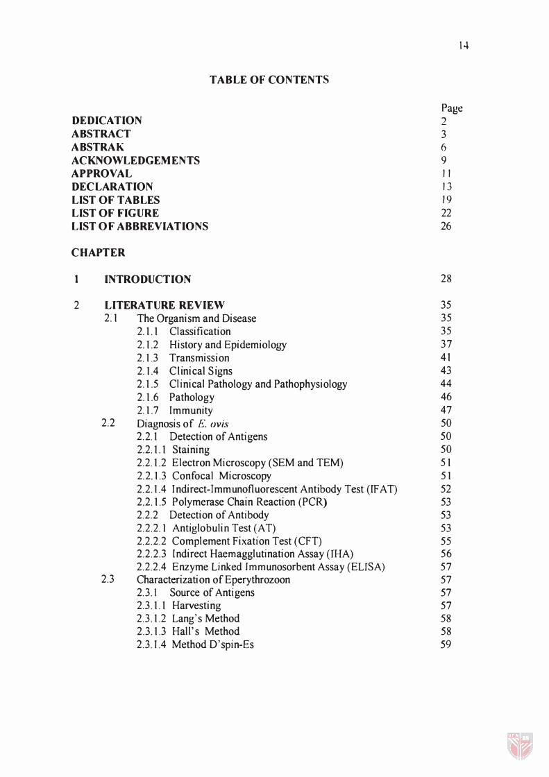
TABLE OF CONTENTS
DEDICATION ABSTRACT A BSTRAK ACKNOWLEDGEMENTS APPROVAL DECLARA TION LIST OF TABLES LIST OF FIGURE LIST OF ABBREVIATIONS
CHAPTER
1
2
INTRODUCTION
LITERATURE REVIEW 2.1 The Organism and Disease
2.1.1 Classification 2.1.2 History and Epidemiolo!,'Y 2.1.3 Transmission 2.1.4 Clinical Signs 2.1.5 Clinical Pathology and Pathophysiology 2.1.6 Pathology 2.1.7 Immunity
2.2 Diagnosis of E. ()vis 2.2.1 Detection of Antigens 2.2.1.1 Staining 2.2.1.2 Electron Microscopy (SEM and TEM) 2.2.1.3 Confocal Microscopy 2.2.1.4 I ndirect-Immunofluorescent Antibody Test (IF AT) 2.2.1.5 Polymerase Chain Reaction (PCR) 2.2.2 Detection of Antibody 2.2.2.1 Antiglobulin Test (AT) 2.2.2.2 Complement Fixation Test (CFT) 2.2.2.3 Indirect Haemagglutination Assay (IHA) 2.2.2.4 Enzyme Linked Tmmunosorbent Assay (ELISA)
2.3 Characterization of Eperythrozoon 2.3. 1 Source of Antigens 2.3 .1.1 Harvesting 2.3 .1.2 Lang's Method 2.3 .1.3 Hal l's Method 2.3 . ] .4 Method D'spin-Es
14
Page ')
3 6 9 11 13 19 22 26
28
35 35 35 37 41 43 44 46 47 50 50 50 51 51 52 53 53 53 55 56 57 57 57 57 58 58 59
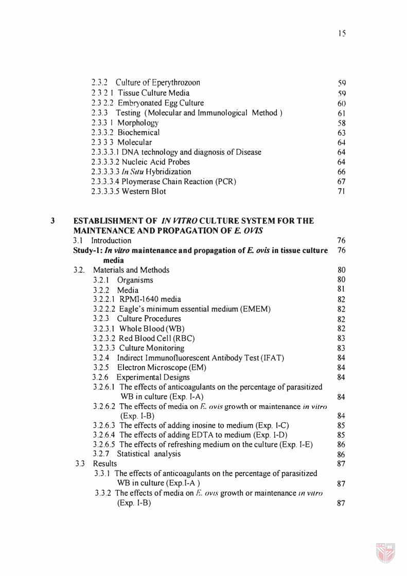
2.3.2 Culture of Eperythrozoon 2 3 2 1 Tissue Culture Media 2.3 2.2 Embryonated Egg Culture 2.3.3 Testing ( Molecular and Immunological Method ) 2.3.3 I Morpholob'Y and Development 2.3.3.2 Biochemical 2.3 3 3 Molecular 2.3.3.3.1 DNA technolob,), and diagnosis of Disease 2.3.3.3.2 Nucleic Acid Probes 2.3.3.3.3 In Situ Hybridization 2.3.3.3.4 Ploymerase Chain Reaction (PCR) 2.3.3.3.5 Western Blot
3 ESTABLISHMENT OF IN VITRO CULTURE SYSTEM FOR THE MAINTENANCE AND PROPAGATION OF E. OVIS
15
59 59 60 61 58 63 64 64 64 66 67 71
3.1 Introduction 76 Study-I: In vitro maintenance and propagation of E. ovis in tissue culture 76
media 3.2. Materials and Methods 80
3.2.1 Organisms 80 3.2.2 Media 81 3.2.2.1 RPMI-1640 media 82 3.2.2.2 Eagle's minimum essential medium (EMEM) 82 3.2.3 Culture Procedures 82 3.2.3.1 Whole Blood (WB) 82 3.2.3.2 Red Blood Cell (RBC) 83 3.2.3.3 Culture Monitoring 83 3.2.4 Indirect Immunofluorescent Antibody Test (IF AT) 84 3.2.5 Electron Microscope (EM) 84 3.2.6 Experimental Designs 84 3.2.6.1 The effects of anticoagulants on the percentage of parasitized
WB in culture (Exp. I-A) 84 3.2.6.2 The effects of media on E. ovis growth or maintenance in vitro
(Exp. I-B) 84 3.2.6.3 The effects of adding inosine to medium (Exp. I-C) 85 3.2.6.4 The effects of adding EDTA to medium (Exp. I-D) 85 3.2.6.5 The effects of refreshing medium on the culture (Exp. I-E) 86 3.2.7 Statistical analysis 86
3.3 Results 87 3.3.1 The effects of anticoagulants on the percentage of parasitized
WB in culture (Exp.I-A ) 87 3.3.2 The effects of media on F;, OV1S growth or maintenance In vlfro
(Exp. I-B) 87
16
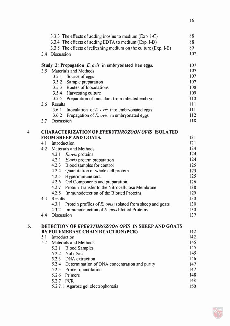
3.3.3 The effects of adding inosine to medium (Exp. I-C) 88 3.3.4 The effects of adding EDTA to medium (Exp. I-D) 88 3.3.5 The etTects of refreshing medium on the culture (Exp. I-E) 89
3.4 Discussion 102
Study 2: Propagation E. ovis in embryonated hen eggs. 107 3.5 Materials and Methods 107
3.5.1 Source of eggs 107 3.5.2 Sample preparation 107 3. 5.3 Routes of Inoculations 108 3.5.4 Harvesting culture 109 3.5.5 Preparation of inoculum from infected embryo 110
3.6 Results 1 11 3.6.1 Inoculation of E OVIS into embryonated eggs 111 3.6.2 Propagation of E. ovis in embryonated eggs 112
3.7 Discussion 118
4. CHARACTERIZATION OF EPERYTHROZOON o VIS ISOLATED FROM SHEEP AND GOATS. 121 4.1 Introduction 121
4.2 Materials and Methods 124
4 .2.1 E.avis proteins 124
4.2.1 E. ovis protein preparation 124 4.2.3 Blood samples for control 125 4.2.4 Quantitation of whole cell protein 125 4.2.5 Hyperimmune sera 125 4.2.6 Gel Components and preparation 126 4.2.7 Protein Transfer to the Nitrocel lulose Membrane 128 4.2.8 Immunodetection of the Blotted Proteins 129
4.3 Results 130 4.3.1 Protein profiles of E. avis isolated from sheep and goats. 130 4.3.2 Immunodetection of E. avis blotted Proteins. 130
4.4 Discussion 137
5. DETECTION OF EPERYTHROZOON OVIS IN SHEEP AND GOATS BY POLYMERASE CHAIN REACTION (PCR) 142 5.1 Introduction 142 5.2 Materials and Methods 145
5.2.1 Blood Samples 145
5.2.2 Yolk Sac 145 5.2.3 DNA extraction 146 5.2.4 Determination of ON A concentration and purity 147 5.2.5 Primer quantitation 147 5.2.6 Primers 148
5.2.7 PCR 148 5.2.7.1 Agarose gel electrophoresis 150
17
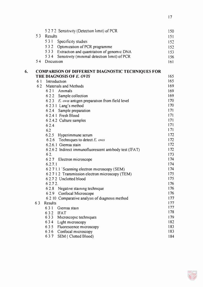
5 2 7 2 Sensitivity (DetectIOn limit) of peR 150 53 Results 151
531 Specificity studies 152 5 3 2 OptimizatIOn of PCR programme 152 5 3 3 ExtractIon and quantltatlOn of genomic DNA 153 5 3 4 SensItivity (minimal detection limit) ofPCR 156
54 DISCUSSion 161
6. COMPARISON OF DIFFE RENT DIAGNOSTIC TECHNIQUES FOR THE DIAGNOSIS OF E. OVIS 165 6 1 Introductton 165
6 2 Matenals and Methods 169 6 2 1 Ammals 169 6 2 2 Sample col lection 169 6 2 3 E. OViS antigen preparatIon from field level 170
6 2 3 1 Lang's method 170
6 2.4 Sample preparation 171
62.4 1 Fresh Blood 171 6 2.4.2 Culture samples 171
62 .4 .2.1 Blood 171
6.2 4 2.2 Yolk sac 171
6.2 . 5 Hyperimmune serum 172 62.6 Techmques to detect E. OVlS 172 6.2.6.1 Giemsa stain 172
62.62 Indirect immunofluorescent antibody test (!FAT) 172 6 2 . 6 3 Polymerase chain reaction (PCR) 173 627 Electron microscope 174 6.2.7 .1 Clotted blood 174 6 2 7 1 .1 'Scanning electron microscopy (SEM) 174 6 2 7 1 2 Transmission electron microscopy (TEM) 17 5 6 2.7 2 Unc10tted blood 17 5 62.7 2 . 1 Scanning electron microscopy (SEM) 176 62.8 Negative stammg techmque 176 62 .9 Confocal Microscope 176 6 2 10 Comparative analYSIS of diagnOSIs method 177
6 3 Results 1 77
6 3 1 Glemsa stam 1 77
6 3 2 IFAT 178
6 3 3 MicroSCOPIc techmques 179 6 3 4 Light microscopy 182 63 5 Fluorescence microscopy 183 636 Confocal microscopy 183 6 3 7 SEM ( Clotted Blood) 184
6.3.8 TEM (Clotted Blood) 6.4 Discussion
7 GENERAL DISCllSSION AND CONCLllSION
BIBLIOGRAPHY
APPENDICES A B C D E F
BIODATA OF THE AUTHOR
184 1 92
195
203
226 227 231 233 238 239 241
244
18
Table
=: I
22
') .., - .)
24
3 1
19
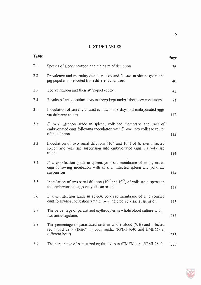
LIST OF T .\BLES
Page
SpecIes of EpelJ1hrozoon and theIr sIte of detectIon 311
Prevalence and mortailty due to [� OVI.\ and f .\u/\ In sheep. goats and pIg populatIOn reported from dIfferent countries 40
Eperythrozoon and their arthropod vector 42
Results of antlglobulms tests m sheep kept under laboratory condItIOns 54
InoculatIOn of serially dtluted E. OVIS mto 8 days old embryonated eggs
via dIfferent routes 113
E. aVIs mfectlOn grade m spleen, yolk sac membrane and liver of embryonated eggs followmg moculatlOn Wlth E. ()VI� mto yolk sac route of moculatlOn 113
33 InoculatIon of two senal ddutlOns (10-2 and ID-l) of E. OVIS Infected spleen and yolk sac suspenSIon mto embryonated eggs VIa yolk sac route 1 14
34 E aVIs mfection grade m spleen, yolk sac membrane of embryonated eggs follo\\lng IncubatIOn Wlth E. OVI.\ mfected spleen and yolk. sac suspenSIon
3 5 InoculatIOn of two senal dIlutIOn (10-2 and 10-3) of yolk sac suspensIOn
114
mto embryonated eggs vIa yolk sac route 1 15
3 6 E. OVIS mfectlOn grade m spleen, yolk sac membrane of embryonated eggs folloWlng mcubatlOn vmh E aVI,\ mfected yolk. sac suspensIOn 115
3 7 The percentage of parasItIzed erythrocytes In whole blood culture WIth two antIcoagulants 235
3 8 The percentage of parasItized cells In whole blood (WB) and mfected red blood cells (IRBC) In both medIa (RPM1-1640 and EMEM) at different hours 235
3 9 The percentage of parasItIzed erythrocytes In rEMEMI and RPMI-1640 =:36
3 10 The percentage of parasitized erythrocytes m mfected red blood cell mcubatlOn wIth reduced Eagle's medium contammg mosme wIth EDTA (rEMEMI-EDTA) and RPMI-I640 medium with EDTA (RPMI-1640-
20
EDTA) 236
3.11 The percentage of parasitized erythrocytes m mfected red blood cell culture with medium refreshment every 12 hours and 24 hours and WIthout changmg medium. 237
4 I Molecular weight of polypeptide bands generated by 10% PAGE of pUrIfied f:. OVIS after staining with coomassle blue. 131
4.2 E. OVIS blotted polypeptide bands immunologically detected by hypenmmune serum agamst antl- E. OVIS 133
4.3 E. OVIS blotted polypeptide bands immunologically detected by nonnal serum. 135
5 1 Oligonucleotide primers used for 16 S rRNA amplification 148
5 2 PCR reaction mixture for the amplification of 16S rRNA of E. OVIS 149
5 3 Detection of E. aVIs by polymerase cham reaction in sheep, goats and 151 yolk sac samples.
5 4 Quantitation of E. aVIs DNA isolated from sheep and goats blood and yolk sac membrane by a spectrophotometer. 155
5 5 Sensitivity of PCR in dIfferent dilution in sheep and goats 156
6 1 Diagnostic techniques for Eperythrozoon antigens from blood samples. 166
62 Sensitivity and spe cificity fonnula 1 73
6 3 DetectIOn of E. OVIS by mdIrect Immunofluorescent antIbody test (IFAT) and Giemsa stam from fresh blood, blood culture and embryonated eggs 178
6 4 Sensitivity and specificity of IFAT and Glemsa stam for the detectIOn of E. aVIs from fresh blood, culture blood and yolk sac membrane 178
6 5 Grade of infection of E. aVIs infected blood by Glemsa and IF AT 179
6 6 DetectIOn of E. aVIs by dIfferent mIcroscoPIc techmques 180
6.7 Sensitivity and specificity of light microscope and tluorescence
21
microscope for the detection of F. OVIS from sheep and goats blood. 1 80
6.8 Sensitivity and specificity of light microscope and confocal microscope for the detection of I�·. OVIS from sheep and goats blood. 1 8 1
6 .9 Sensitivity and specificity of light microscope and TEM for the detection of I�·. ()vis from sheep and goats blood. 18 1
6.10 Sensitivity and specificity of light microscope and SEM for the detection of E. ()vis from sheep and goats blood. 1 8 1
6.11 Comparison of the results of IFAT and Giemsa stain in fresh blood, culture and yolk sac membrane. 239
6.12 Comparison of the results of light mIcroscope and fluorescence microscope for the detection of E. ()vis. 239
6.13 Comparison of the results of light microscope and confocal microscope for the detection of E. ()vis. 240
6.14 Comparison of the results of light microscope and TEM and SEM for the detection of E. ()vis. 240
22
LIST OF FIGURES
Figure Page
3.1 The percentage of parasitized erythrocytes in whole blood culture wtth two anticoagulants. Significant difference observed (p < 0.05) between heparin and EDT A anticoagulant in whole blood incubation over the 408 hours of observations. 91
3.2 The percentage of parasitized cells in whole blood (WB) and infected red blood cells (lRBC) in both media (RPMI-1640 and EMEM) at different hours. Significant difference (p < 0.05) observed between RPMI-1640 and EMEM in WB and IRBC incubation over the 408 hours of observations. 92
3.3 The percentage of parasitized erythrocytes in infected red blood cell culture with reduced Eagle's medium containing inosine (rEMEMl) and RPMI-1640 medium without inosine. Significant difference (p < 0.05) observed between reduced Eagle's medium containing inosine and RPMI-1640 medium without inosine over the 408 hours of observations. 93
3.4 The percentage of parasitized erythrocytes in infected red blood cell incubation with reduced Eagle's medium containing inosine with EDTA (rEMEMI - EDTA) and RPMI-1640 medium with EDTA (RPMI-1640-EDTA). Significant difference (p < 0.05) observed between rEMEMI - EDTA and RPMI-1640-EDTA in infected red blood cell incubation over the 408 hours of observations. 94
3.5 The percentage of parasitized erythrocytes in infected red blood cell culture with medium refreshment every 12hours and 24 hours, and without changing medium. Significant d ifference (p < 0.05) observed between no change and 12 hours and between no change and 24 hours over the 408 hours of observations. 95
LIST OF PLATES
Plate
3. 1 Giemsa stained smears of the I�·. OVIS (arrow) from culture containing
23
Page
RPM1- 1640 medium in 12 hours (x 100). 96
3.2 Giemsa stained smears of the F. OVIS (arrow) from culture containing EMEM medium in 12 hours (x 100). 96
3.3 Giemsa stained smears of the E. ()vis (arrow) from culture containing RPM1- 1640 medium in 24 hours (x 100). 97
3.4 Giemsa stained smears of the E. OVIS (arrow) from culture containing EMEM medium in 24 hours (x 100). 97
3.5 Giemsa stained smears of the E. ()vis (arrow) from culture containing RPMI-1640 medium in 48 hours (x 100). 98
3.6 Giemsa stained smears of the E. ovis (arrow) from culture containing EMEM medium in 48 hours (x 100). 98
3.7 Giemsa stained smears of the E. ovis (arrow) from culture containing RPMI-1640 medium in 156 hours (x 100). 99
3.8 Giemsa stained smears of the E. avis (arrow) from culture containing EMEM medium in 156 hours (x 100). 99
3.9 Giemsa stained smears of the E. ovis (arrow) from culture containing EMEM medium in 408 hours (x 100). 100
3.10 Electron micrographs of E. avis from culture (TEM x 40,000) 100
3.1 1 Presence of green fluorescence color (arrow) of E. ovis on the surface of infected culture red blood cell by indirect immuno-fluorescent antibody test (IFAT) (x 400). .. 101
3. 12 Presence of E. ()vis (arrow) in yolk sac membrane of embryonated egg culture by Giemsa stain (x 100). 116
3.13 Presence of green fluorescence color (arrow) in yolk sac membrane of embryonated egg culture by indirect immuno-fluorescent antibody test ( IFAT) (x400) 116