UNIVERSITI PUTRA MALAYSIA CLONING OF … tesis yang dikemukakan kepada Senat Universiti Putra...
25
UNIVERSITI PUTRA MALAYSIA CLONING OF CHICKEN ANAEMIA VIRUS (CUX-I) VPI GENE INTO A LACTOCOCCUS EXPRESSION VECTOR pMG36e NG PERK TSONG FSMB 1999 6
-
Upload
truonghanh -
Category
Documents
-
view
216 -
download
0
Transcript of UNIVERSITI PUTRA MALAYSIA CLONING OF … tesis yang dikemukakan kepada Senat Universiti Putra...
UNIVERSITI PUTRA MALAYSIA
CLONING OF CHICKEN ANAEMIA VIRUS (CUX-I) VPI GENE INTO A LACTOCOCCUS EXPRESSION VECTOR pMG36e
NG PERK TSONG
FSMB 1999 6
CLONING OF CHICKEN ANAEMIA VIRUS (CUX-I) VPI GENE INTO A LACTOCOCCUS EXPRESSION VECTOR pMG36e
By
NG PERK TSONG
Thesis Submitted in Fulfilment of the Requirements for the Degree of Master of Science in the
Faculty of Food Science and Biotechnology Universiti Putra Malaysia
August 1999
Abstract of thesis presented to the Senate ofUniversiti Putra Malaysia in fulfilment of the requirements for the degree of Master of Science
CLONING: OF CmCKEN ANAEMIA VIRUS (CUX-l) VPl GENE INTO A LACTOCOCCUS EXPRESSION VECTOR pMG36e
By
NG PERK TSONG
August 1999
Chairperson: Raha Abdul Rahim, Ph.D.
Faculty: Food Science and Biotechnology
Lactococcus lac/is is a non-pathogenic and non-colonising bacterium, which is being
developed as a vaccine delivery vehicle for immunisation by mucosal routes. To
determine whether lactococci can express the Chicken Anaemia Virus capsid protein
(VPI ) in immunogenic fonn, we have constructed a recombinant DNA, which
constitutively expresses the VPI as N-tenninal fusion protein with Open Reading
Frame-32 under the control of lactococci promoter P32 in vector pMG36e.
Chicken anaemia virus (CAV) is a member of the newly categorised Circoviridae, a
family of circular negative single-stranded DNA viruses. CAY VPI gene ( 1 .4 kb)
was amplified by PCR from CA V genome which was isolated from infected Marek's
disease virus transfonned lymphoblastoid cell line (MDCC-MSB 1 ) cell lysate and
cloned into pCR®2 . I -TOPO vector for sequencing. The sequenced VPI gene,
lacking its own promoter, was cloned into the lactococcal expression vector
11
pMG36e and transformed into E. coli JM1 09 as the intermediate host. The
recombinant plasmid was sub-cloned into L. lactis MG 1 363 by electroporation.
Several independent verifying methods such as sequencing, Southern hybridisation
and restriction enzymes analysis have confirmed the insertion of VPl gene into
pMG36e. The complete sequence of VPI gene has 99.65% homology compared to
the published CA V Cux-l VPl gene. A total of 5 base difference was seen to result
in change of 3 amino acids sequence in the polypeptide chain. The expression of
fusion protein was studied until transcriptional level . Transcription of fusion gene
from strains of recombinant L. lactis MG 1363 were analysed by Northern
hybridisation. Northern hybridisation has detected transcription of fusion gene in
recombinant L. iactis MG1363 carrying pMG36e-VPl and produced mRNA with the
size of 1.4 kb.
On the basis of these results, it is concluded that the recombinant DNA pMG36e was
successfully constructed in bacteria E. coli JM1 09 and subcloned into L. lactis
MG1363. The recombinant DNA in L. lactis MG1 363 is capable to express the
fusion protein up to transcriptional level. Therefore, recombinant L. lactis MG 1 363
can be used as a potential vaccine delivery system against CA V infections.
111
Abstrak tesis yang dikemukakan kepada Senat Universiti Putra Malaysia sebagai memenuhi keperluan untuk ijazah Master Sains.
PENGKLONAN GEN VPl VIRUS ANAEMIA A YAM (CUX-l) KE DALAM SATUVEKTOR PENGEKPRESLACTOCOCCUSpMG3�
Oleh
NG PERK TSONG
Ogos 1999
Pengerusi: Raha Abdul Rahim, Ph.D.
Fakulti: Sains Makanan dan Bioteknologi
LaclococcuS lactis ialah sejenis bacteria yang bukan patogenik dan tidak
mengkoloni yang telah direka sebagai agen untuk pengangkutan vaksin untuk tujuan
immunisasi melalui kaedah mucosal. DNA rekombinan yang mengekspres VPI
sebagai terminal N protein fusion dengan Open Reading Frame-32 di bawah
pengawalan promoter P32 di dalam vektor pMG36e telah dibina untuk menentukan
sarna ada lactococci boleh mengekspres protein kapsid (VPI ) yang immunogenik.
Virus anemia ayam (CAV) adalah ahli kepada keluarga baru yang dinamakan
Circoviridae, iaitu kumpulan yang mempunyai DNA bulatan negatif satu rantai .
Gen VPI ( 1 .4 kb) telah diamplifikasikan melalui PCR daripada genom CAY yang
diekstrak dari sel MDCC-MSB I yang telah dijangkiti oleh CA V dan diklonkan ke
dalam vektor pCR®2. l -TOPO untuk penentuan jujukan DNA. Gen VPI tanpa
IV
PERPUSTAKAAN JNIVERSlTl PUTRA MALAYSIA
promoter telah dimasukkan ke dalam vektor pengekpres pMG36e dan ditransfom ke
dalam E. coli JM109 sebagai perumah perantara. DNA recombinan ini kemudian
dipindahkan ke dalam bakteria L. lactis MG 1 363 dengan kaedah electroporation.
Beberapa kaedah pengesahan termasuk penentuan jujukan DNA, analisis dengan
enzim pembatas dan penghibridan Southern telah menunjukkan bahawa gen VPl
telah berjaya dimasukkan ke dalam vektor pMG36e. Keseluruhan penentuan jujukan
DNA menunjukkan gen VPl mempunyai homologi 99.65% berbanding dengan
jujukan DNA gen VPl CAY Cux-l yang telah diterbit. Sejumlah 5 perbezaan bes
dalam jujukan DNA telah membawa perubahan kepada 3 jujukan asid amino di
dalam rantai polipeptida. Kajian terhadap pengekspresan protein fusion telah
mencapai ke tahap transkripsi. Transkripsi gen fusion daripada strain L. lactis
MG1363 rekombinan telah dianalisis dengan penghibridan Northern. Penghibridan
Northern menunjukkan bahawa transkripsi gen fusion telah belaku di dalam strain L.
lactis MG1 363 yang mengandungi pMG36e-VPl dan menghasilkan mRNA bersaiz
1 .4 kb.
Berdasarkan hasil kajian yang diperolehi, ia boleh disimpulkan bahawa DNA
rekombinan telah berjaya dibina dalam bakteria E. coli JMI09 and disubklon ke
dalam L. iactis MG1 363. DNA rekombinan di dalam L. lac/is MG1 363 berupaya
mengekspres protein fusion sehingga tahap transkripsi. Oleh itu, L. lactis MG 1 363
rekombinan berpotensi untuk digunakan sebagai sistem pengangkutan vaksin bagi
mencegah jangkitan CA V.
v
ACKNOWLEDGEMENTS
The author wishes to express his deepest gratitude to supervisors, Dr. Raha Abdul
Rahim of Biotechnology Department, Associate Professor Dr. Khatijah Mohd.
Yusoff and Associate Professor Dr. Abdullah Sipat of Biochemistry and
Microbiology Department for their invaluable advises and guidance throughout the
course of this project.
The author wishes to express his thanks to Miss Ernie Eileen Rizlan Ross, Miss
Norfilza Ahmat, Meilina Ong, Kak Liza and all members of Microbial Molecular
Laboratory, Genetic Laboratory and ATCL for their support and co-operation
throughout the project.
Lastly, the author wishes to thank all friends who have contributed their idea and
help to him.
VI
I certify that an Examination Committee met on 19th August, 1999, to conduct the final examination of Ng Perk Tsong, on his Master of Science thesis entitled "Cloning Of Chicken Anaemia Virus (Cux-l) VPl Gene into A Lactococcus
Expression Vector pMG36e" in accordance with Universiti Pertanian Malaysia (Higher Degree) Act 1980 and Universiti Pertanian Malaysia (Higher Degree) Regulations 1981. The Committee recommends that the candidate be awarded the relevant degree. Members of the Examination Committee are as follows:
RAHA ABDUL RAHIM, Ph.D. Faculty of Food Science and Biotechnology Universiti Putra Malaysia (Chairman)
KHATIJAH MOHO. YUSOFF, Ph.D. Associate Professor Faculty of Science and Environmental Studies Universiti Putra Malaysia (Member)
ABDULLAH SIP AT, Ph.D. Associate Professor Faculty of Science and Environmental Studies Universiti Putra Malaysia (Member)
TAN SIANG HEE, Ph.D. Faculty of Food Science and Biotechnology Universiti Putra Malaysia (Independent Examiner)
MOHO ALI MOHA YIDIN, Ph.D, Professor/ Deputy Dean of Graduate School) Universiti Putra Malaysia
Date: 24 JAN 2000
VI1
This thesis was submitted to the Senate of Universiti Putra Malaysia and was accepted as fulfilment of the requirements for the degree of Master of Science.
KAMIS A WANG, Ph.D, Associate Professor Dean of Graduate School, Universiti Putra Malaysia
Date: 11 MAY 2000
VlIl
DECLARATION
I hereby declare that the thesis is based on my original work except for quotations and citations, which have been duly acknowledged. I also declare that it has not been previously or concurrently submitted for any other degree at UPM or other institutions.
Signed
Candidate. Ng Perk Tsong 21 sl February 2000
IX
TABLE OF CONTENTS
Page
ABSTRACT . . . . . . . . . . . . . . . . . . . . . . . . . . . . . . . . . . . . . . . . . . . . . . . . . . . . . . . . . . . . . . . . . . . . . 11 ABSTRAK . . . . . . . . . . . . . . . . . . . . . . . . . . . . . . . . . . . . . . . . . . . . . . . . . . . . . . . . . . . . . . . . . . . . . . . IV ACKNOWLEDGMENTS . . . . . . . . . . . . . . . . . . . . . . . . . . . . . . . . . . . . . . . . .. . . . .. . . . .. VI APPROVAL SHEETS . . . . . , ... . , . ... . , . ... ... ... ... .. , '" ., . ... ... ... ... '" ... V11 DECLARATION FORM . . . . . . . . . . . . . . . . . . . . . . . . . . . . . . . . . . . . . . . . . . . . . . . . . . . . . IX LIST OF TABLES... . . . ... ... ... ... ... ... ... ... ... ... ... ... ... ... ... ... ... .... Xlll LIST OF FIGURES . . . .. . . . , ... .. , .... , . ... '" ... ... ... ... .. , .... , . ... ...... '" XIV LIST OF PLATES . . . . . . . . . . . . . . . . . . . . . . . . . . . . . . . . . . . . . . . . . . . . . . . . . . . . . . . . . . . . . xv LIST OF ABBREVIATIONS . . . . . . . . . . . . . . . . . . . . . . . . . . . . . , ... . , . ... . ,. ... ... XVI
CHAPTER
I INTRODUCTION . . . . . , . . . . . , . . . . ,. '" . . . . . . . . . . . . '" . . , . . . . , . . . . '" . . . 1 Objectives . . . . . . . . . . . . . . . . . . . . . . . . . . . . . . . . . . . . . . . . . . . . . . . . . . . . . . . . . . . . . . . . . . 3
n LITERATURE REVIEW . . . . . . . . . . . . . . . . . . . . . . . . . . . . . . '" . . . . . . . . . . . . 4 Chicken Anaemia Virus (CAV) . . . . , . . . . . . . . . . . . . . . . . . . . , . . . . '" . . . . . . . 4
Introduction . . . . . . . . . . . . . . . . . . . . . . . . . . . . . . . . . . . . . . . . . . . . . . . . . . . . . . . 4 Genomic Organisation and Gene Products of CA V (Cux-l) . . , . . . . , . . . . .. . . . .. . . '" . . . . . , . . .. , . . .. . . . . . . . . . '" . . . . . , . . . . 6
Molecular Cloning of CA V Genome or Genes . . . . . . . . , . . . . . . . . . '" . . . 9"i' Lactic Acid Bacteria and Lactococcus . . , . . . . . . . . .. . . . .. . . . . . . . . . . . . . . . . 1 1
Introduction . . . . . . . . . . . . . . . . . . . . . . . . . . . . . . . . . . . . . . . . . . . . . . . . . . , ... ] 1 Gene Expression Vectors for Lactococci . . . . . . . . . . . . . . . . . , . . . 12-1 Plasmid Replication and Stability in L. lactis ' " . . . . . . . . . . . . 14
Gene Transfer in Lactococci . . . . . . . . . . . . . . . . . . .. . . . . . . . . . . . . . . . . . . . . . . . . 18 Introduction . . . . . . . . . . . . . . . . . . . . . . . . . . . .. . . . . . . . . . . . . . .. . . . . . . . . . . 18 Transformation of L. lactis via Electroporation . . . . . . . . . . . . 1 8
Lactic Acid Bacteria as Vaccine Delivery Vehicle . . . . ,. '" . . . . . . '" 25 Introduction . . . . . . . . . . . . . . . . . . . . . . . . . . . . . . . . . . . . . . . . . .. . . . . . . . . . . 25 Antigens Expression in L. lactis . . . . . . . . . . . . . . . . . . . . . . . . . . . . . . 27
m MATERIAL AND METHODS . . . . . . . . , '" . . . . . . '" . . . '" . . . . . . . . . . . 30 Bacterial Strains, Plasmids and Media . . . . . . . . . . . . . . . . . . . . . . . . . . . . . . . 30 Isolation of Viral DNA '" . , . . . . . . . . . . . . . . . , . . . . . . . . . . , . . . . . . . . . . . . . . . , . . 30 Preparation of Competent Cells '" . . . '" ., . . . . . . . . . . , . . . . . . . . . . ,. . . . . . . 32
E. coli JM109 and XLI-blue MRF' . . . . . . ... . . . .. , . . . . . . . .. . . . 32 L. lac/is MG 1363 ., . . . . . . . . . . . . . . . . . . . . . , . . . . . , . . . . . . . . . '" . . . . . . 3 3
X
Page
Miniprep Plasmid Isolation by Alkaline Lysis Method (Birboim and Doly, 1979) . . . ... . . . . . . . . . . . . . . . . . . . . . . .. _ . . . . . . . . . . . . ,. 33
E. coli Top 10, JM109 and XLI-blue MRF' . . . . . . . . . .. . . . . . 33 L. lactis MG 1363 ...... ...... ... ... ... ......... ................. 35
Agarose Gel Electrophoresis ......... .. , ... ... ... ... ... ... ... ... ... .... 35 Restriction Enzymes Digestion . .. ... ... ... ... . . . . .. ... ... ... . .. ... ... 36 Polymerase Chain Reaction (PCR) ...... ... ... ......... ...... ...... ... 37
Amplification of CA V (Cux-l) VPl Gene ... ... ... ...... .. 37 Verification of Recombinant DNA .. , .............. , ... ...... 38
Cloning of CAV (Cux-l) VPl Gene .. , ...... '" ... .. , ... ...... ... .... 39 Cloning ofVPI Gene into pCR®2.1-TOPO ....... '" ... .... 39 Subcloning ofVPl Gene into pMG36e ... . , . ...... ...... .... 40
Transformation of Bacteria ...... ... ............ ................... ..... 41 Transformation of E. coli Top 10, JM109 and XLI-blue MRF ' . . . .. . . . . . . , ... ...... ... ......... '" ..... , ... ... .. 41 Transformation of pMG36e and Recombinant DNA into L. lac/is MG 1363 ., . .................... , ...... . , . ..... , ...... . ,. 42
Selection of Recombinants ... ...... ... .. , ... '" ........ , ... ............. 42 Sequencing ofVPl Gene ... ... ... ............... ... ... ...... ...... ..... 43
Sequencing ofVPl Gene in Plasmid pCR®2.1 ... ... ... .... 43 Sequencing ofVPl Gene in Plasmid pMG36e ............ ,. 44
Southern Hybridisation (Southern, 1975) ......... ... ................ 45 Preparation of DIG-labelled Probe ... ... ..... , ... ...... '" .... 45 Quantification of DIG-labelled Probe ... .... , . ...... ... .... ,. 45 Transfer of DNA to Nylon Membrane ... ............ ........ 48 Hybridisation Procedure ... ......... ... ......... ......... ...... 49 Detection Procedure ...... ... .................. ......... ........ 50
Northern Hybridisation . . . . . . . . . . . . . . . . . . . . . . . . . . . . . . . . . . . . . . . . . . . . . . . . . . 51 Total RNA Extraction ...... ...... .............. , ...... ...... .... 51 Formaldehyde Agarose Gel Electrophoresis ... ... ... ... ..... 52 Preparation of DIG-labelled DNA Probe, RNA Marker and Transfer of Total RNA to Nylon Membrane ........... 53 Membrane Hybridisation and Immunological Detection .. 53
IV RESULTS AND DISCUCCION . . . . . . . .. . . . . . . . . . . . . .. . . . . . . . . . . . . . 54 Isolation of Viral DNA and PCR Amplification of VPl Gene .... 54 Cloning and Sequencing of PCR Product in pCRe2.1-TOPO ...... 56 Cloning ofVPl Gene into pMG36e and Transformation into E. coli JM109 .. , ... ......... .. , ... ...... ... .. , ... '" .................. 64 Transformation of pMG36e-VPl DNA into L. lactis MG1363 . _ . 67 Partial Verification of Recombinant DNA (pMG36e-VP1) by Restriction Enzymes Analysis .............. , .............. , ... ..... 69
Xl
Page
Verification of Recombinant DNA (pMG36e-VPl) by Other Methods . . . . . . . . . . . . . . . . . . . . . . . . . . . . . . . . . . . . . . . . . . . . . . . . . . . . . . . . . . . , 72
PCR Technique . . . . . . . . . . . . . . . . . . . . . . . . . . . . . . . . . . . . . . . . . . . . . . . . . 72 Southern Hybridisation . . . . . . . . . . . . . . . . . . . , . . . . . , . . . . . . , . . . . . . . 74 Sequencing ofVPl Gene in pMG36e . . . . . . . . . . . . . . . . . . . . . . .. 77
Expression of VP 1 Gene in L. lactis MG 1363 . . . . . . . . . . . . . . . . . . . . . . . . 78 Total RNA Extraction . . . . . . . . . . . . . . . . . . . . . . . . . . . . . . . . . . . . . . . . . . 78 Northern Hybridisation . . . . . . . . . . . . . . . . . . . .. . . . . . . . . . . . . . . . . . . . . 79
V GENERAL DISCUSSION . . . . . . . . . . . . . . . . . . . . . . . . . . . . . . . . . . . . . . . . . . . . 82
VI CONCLUSIONS . . . . . . . . . '" . . . . . . . . . . . . . . . . ,. . . . . . . . . . . . . . . . . . . . . . . . . . . 87
REFERENCES . . . . . . . . . . . . . . . . . . . . . . . . . . . . . . . . . . . . . . . . . . . . . . . . . . . . . . . . . . . . . . . . . . . 89
APPENDICES . . . . . . . . , . . . . . . . . . . . . . . . . . . . . . . . . . . . . . . . . . . . . . . . . . . . . . . . . . . . . . . . . . . . . 99
Appendix A: Composition of Media and Solutions . . . . . . . . . . . . . . . . . , 100 Appendix AI: Composition of Media and Antibiotic Stock
Solutions .. . . . . . . . . . . .. . . . . . . . . . . . . . . , . . . . . . , . . . . . . . . . . . . , 100 Appendix A2: Solutions for Alkaline Lysis and Agarose
Gel Electrophoresis . . . . . . . . . . . . . . . . . . . . . . . . . . . . . . . . . . . . . 102 Appendix A3: Solutions for Competent Cell Preparation . . . . . . . . . 103 Appendix A4: Solutions and Buffers for Southern and Northern
Hybrididation . . , . . . . . . '" . . . . , . . . . . . , . . . . . . . . . . . . ,. . . . . . 104 Appendix B: Sizes of DNA Markers . . . . . . . . , . . . . . , . . . . . . . . . . . . . . . . . . . 106 Appendix C: Comparison between VPl Gene Automated
Sequencing Results (upper sequence) and Published CAV Cux-l DNA Sequences (lower sequence) . . . . 107
BIODATA OF AUTHOR . . . . . . . . . . . . . . . . . . . . . . . . . . . . . . . . . . . . . . . . . . . . . . . . . . . 109
XII
LIST OF TABLES
Table Page
1 Transformation of L. lactis . . . . . . . . . . . . . . . . . . . . . . . . . . . . . . . . . . . . . . . . . . . . . 20
2 Antigens Expressed in L. lactis . . . . . . . . . . . . '" . . . . . . . . . . . . . . . . . , . . . . . . . 29
3 Bacterial Strains and Plasmids . . . . . . . . . . . . . , . . . . . . . . . . . . . . . . . . . . , . . . . . . . 31
4 Characteristics of Restriction Enzymes . . , . . . . . . . . . '" . , . . . . . . , . . . . . . . . 37
5 Characteristics of PCR Primers '" . . . . . . . . . . . . . . . . . . . . . . . . . . . . . , . . . . . . . 38
6 Characteristics of Sequencing Primers . . . . . . . . . . , . . . . . . , . . . . . . . . . . . . . . 39
7 Components of 5% (v/v) Long Ranger Sequencing Gel . . . . , . . . . . . , 44
8 Dilution of DIG-labelled Probe . . . . . . . . . . , . . . . . . , '" . . . '" . . . . . . . . . . . , 46
9 Teststrips Processing Sequence '" . , . . . . . . . . . . . . . '" . . . . , . . . . . . . . . . . . . . . 47
XllI
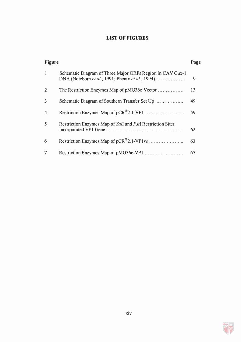
LIST OF FIGURES
Figure
1 Schematic Diagram of Three Major ORFs Region in CAV Cux-l
Page
DNA (Noteborn et aI., 1991; Phenix et a!., 1994) ... '" ... ... ... ... 9
2 The Restriction Enzymes Map ofpMG36e Vector .. , ... ... ... .... 13
3 Schematic Diagram of Southern Transfer Set Up ... ... ... ... ..... 49
4 Restriction Enzymes Map [email protected].. .... ..... , ... ... ... .... 59
5 Restriction Enzymes Map of SalI and PstI Restriction Sites Incorporated VP 1 Gene . . . . . . . . . . . . . . . . . . . . . . . . . . . . . . . . . . . . . . . . . . . . . . . 62
6 RestrictionEnzymes Map [email protected] ... ... ................ 63
7 Restriction Enzymes Map ofpMG36e-VP1 ...... .... ,. ... ... ... ... 67
XIV
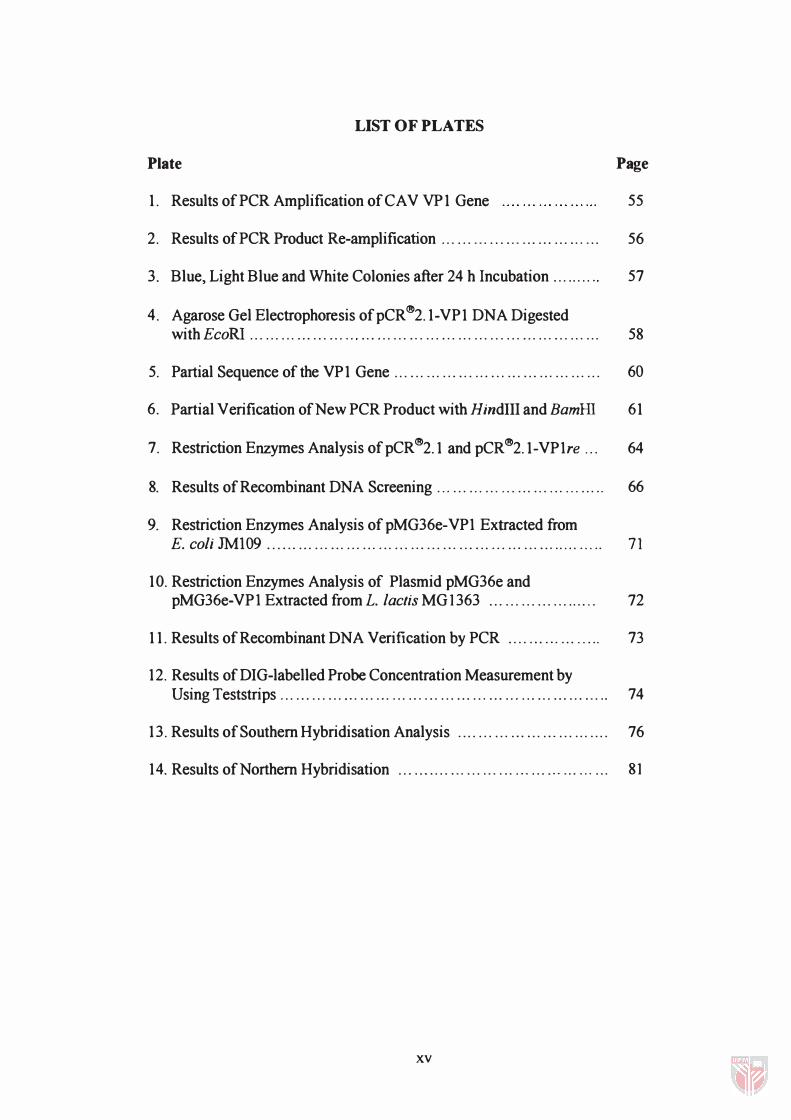
LIST OF PLATES
Plate
1. Results of PCR Amplification of CA V VP 1 Gene
Page
55
2. Results of PCR Product Re-amplification . . . .. . . . . ...... . .. ... ..... , . .. 56
3. Blue, Light Blue and White Colonies after 24 h Incubation .. . ... . . " 57
4. Agarose Gel Electrophoresis of pCR �2.1-VP 1 DNA Digested with EcoRI . .. . . . . .. . .. . .. ... '" .. . . . . . . . . , . . . . . , . . . . . , . . . . . . , .. . . . , .. . . . , . . . 58
5. Partial Sequence of the VP 1 Gene .. . .. . . .. . , . .. . . , . .. . ... ... . . , . . . . . , . . . 60
6. Partial V erification of New PCR Product with HindllI and BamHI 61
7. Restriction Enzymes Analysis of pCR�2.1 and pCR�2.1-VPlre .. . 64
8. Results of Recombinant DNA Screening . .. . . . . .. . . . '" ... . .. ... ... ... .. 66
9. Restriction Enzymes Analysis of pMG36e-VPl Extracted from E. coli JMI09 ...... . .. .. . ... . . . ... . .. ... . , . . . . . , . .. ... . . . . .. , ... .. , .. ... . .... 71
10. Restriction Enzymes Analysis of Plasmid pMG36e and pMG36e-VPl Extracted from L. lac/is MG 1363 ..... . . . . ... . . ... .... 72
11. Results of Recombinant DNA Verification by PCR .. . . . .. . ,. . .. ..... 73
12. Results of DIG-labelled Probe Concentration Measurement by Using Teststrips ... ......... . , . ... ... . .. .. , . .. .. . ... .. , ... ... . .. .. . ... ... ... .. 74
13. Results of Southern Hybridisation Analysis . ... .. . ... ... ... ... ... . . . . ... 76
14. Results of Northern Hybridisation ... . , . . . .. ... . . . . . . . .. . , . ... ' " . . . ' " .. . 8 1
xv
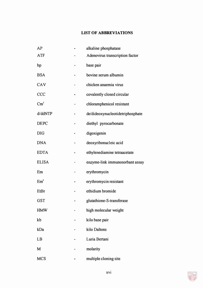
LIST OF ABBREVIATIONS
AP alkaline phosphatase
ATF Adenovirus transcription factor
bp base pair
BSA bovine serum albumin
CAY chicken anaemia virus
CCC covalently closed circular
Cmf chloramphenicol resistant
d/ddNTP de/dideoxynucleotidetriphosphate
DEPC diethyl pyrocarbonate
DIG digoxigenin
DNA deoxyribonucleic acid
EDTA ethylenediamine tetraacetate
ELISA enzyme-link immunosorbant assay
Em erythromycin
Emf erythromycin resistant
EtBr ethidium bromide
GST glutathione-S-transferase
HMW high molecular weight
kb kilo base pair
kDa kilo Daltons
LB Luria Bertani
M molarity
MCS multiple cloning site
XVI
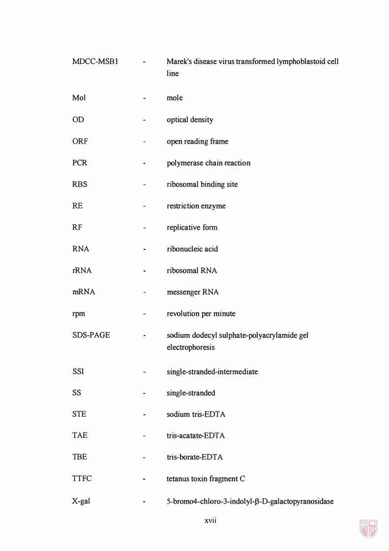
MDCC-MSBI
Mol
OD
ORF
PCR
RBS
RE
RF
RNA
rRNA
mRNA
rpm
SDS-PAGE
SSI
SS
STE
TAE
TBE
TTFC
X-gal
Marek's disease virus transformed lymphoblastoid cell
line
mole
optical density
open reading frame
polymerase chain reaction
ribosomal binding site
restriction enzyme
replicative form
ribonucleic acid
ribosomal RNA
messenger RNA
revolution per minute
sodium dodecyl sulphate-polyacrylamide gel
electrophoresis
single-stranded-intermediate
single-stranded
sodium tris-EDTA
tris-acatate-EDT A
tris-borate-EDT A
tetanus toxin fragment C
5-brom04-chloro-3-indolyl-p-D-galactopyranosidase
XVll
CHAPTER I
INTRODUCTION
Chicken anaemia virus (CA V) is a member of the newly categorised family
Circoviridae. CA V is a small icosahedral shaped DNA virus. The virus causes a
syndrome in chicken characterised by aplastic anaemia, lymphoid depletion,
subcutaneous and intramuscular haemorrhages and increased mortality (Yuasa et
ai. , 1979). It also causes the destruction of the erythroblastoid cells in bone
marrow and depletion of cortical CD4+ lymphocytes in the thymus, resulting in
acute anaemia and immunodeficiency (Jeurissen et ai. , 1989).
Current vaccines for CA V are based on either live attenuated or inactivated CA V
(Steenhuisen et aI. , 1994); however, there are drawbacks in each approach. The
use of attenuated viral vaccines carries the risk that the virus is not truly
attenuated. Consequently, the chickens may become infected by CA V. Such
infections are often asymptomatic and result in lower growth and weight gain
rates.
Several researchers have studied cloning and sequencing of genomic DNA from
CAY (Claessens et aI. , 1991; Noteborn et aI. , 1991; Meehan et aI. , 1992). Gene
sequences have been isolated that could be used to make a subunit vaccine
against CA V. Amplified CA V genes were cloned into plasmids and transformed
to suitable host such as bacteria and baculovirus (pallister, 1994; Kato et al. ,
1995; Koch et aI. , 1995). The expression of the cloned gene(s) was determined
by screening of the fusion protein. Since subunit vaccines utilise recombinantly
expressed proteins of the virus and not the virus itself, the problem of infections
inherent in the use of attenuated viruses as vaccines can be overcomed.
Furthermore, manufacture of recombinant proteins for use as subunit vaccines are
less expensive than the culture and inactivation of the viruses used in inactivated
viral vaccines.
Lactococcus is one of the important groups of lactic acid bacteria. It is a Gram
positive cocci and produces lactic acid during fermentation process. Lactococcus
consists of two major subspecies: L. lactis subsp. lactis and L. lactis subsp.
cremoris. This bacterium has importance for major economic activities,
primarily their use in a variety of dairy fermentation such as cheese production.
Much effort has been put into the development of methods to modify these
microorganisms genetically. Transformation systems were developed and
special-purpose cloning vectors became available (Kok , 1991).
Increasing knowledge of the molecular biology of the lactic acid bacteria creates
the interest in the development of alternative bacterial vaccine vectors based on
2
the harmless and non-pathogenic bacteria as cloning and antigen vehicles. Such a
system would be particularly suited for mucosal immunisation of geriatric and
paediatric populations. In contrast to conventional vaccines, which are largely
derived from attenuated pathogens such as Salmonella and BCG (Dougan et al. ,
1993; Stover et al. , 1993), the lactic acid bacteria are generally regarded as safe
organisms making them particularly attractive for use as antigen delivery
vehicles. L. lactis was able to produce high levels of heterogeneous antigen
expression in vitro prior to inoculation of the bacteria (Norton et al. , 1995).
Therefore this system does not require in vivo expression of the antigen in the
host. Furthermore L. lactis has a low innate immunogenicity and it is possible to
carry out sequential oral inoculations of this bacterium without inducing a
significant increase in the antibacterial antibody response, thus reducing the risk
of interference from antibacterial antibodies (Norton et al. , 1995).
Objectives
The objective of this study is to clone the VP l gene of the CAY Cux-l strain into
the lactococcal expression vector pMG36e. The VPI gene is first amplified by
the polymerase chain reaction and then cloned into the pCR®2.1 cloning vector
for product verification. The VPI gene is then subcloned in-frame with ORF-32
into pMG36e and transformed into L. lactis MG1363. The fusion gene
expression in L. lactis MG 1363 is examined using Northern hybridisation.
3
CHAPTERll
LITERATURE REVIEW
Chicken Anaemia Virus (CA V)
Introduction
CA V is a member of the newly categorised Circoviridae, a family of circular
negative single-stranded DNA viruses that also includes the porcine circovirus
and the beak and feather disease virus of parrots (Studdert, 1993). These
viruses are grouped together on the basis of a common geliome form, but there
were no similarity in amino acid composition, open reading frame (ORF)
arrangement, nor transcriptional machinery have been identified. The virion is
a sma)), icosahedral DNA virus with an average diameter of 25 nm (Goryo el
al. , 1987). It is resistant to treatments with chloroform, ether, pH 3, heating at
80°C for 5 min and can be cultured in Marek's disease virus transformed
chicken lymphoblastoid cell line (MDCC-MSB 1 ).
PERPUSTAKAAN JNIVERSITI PUfRA MALAY§1A
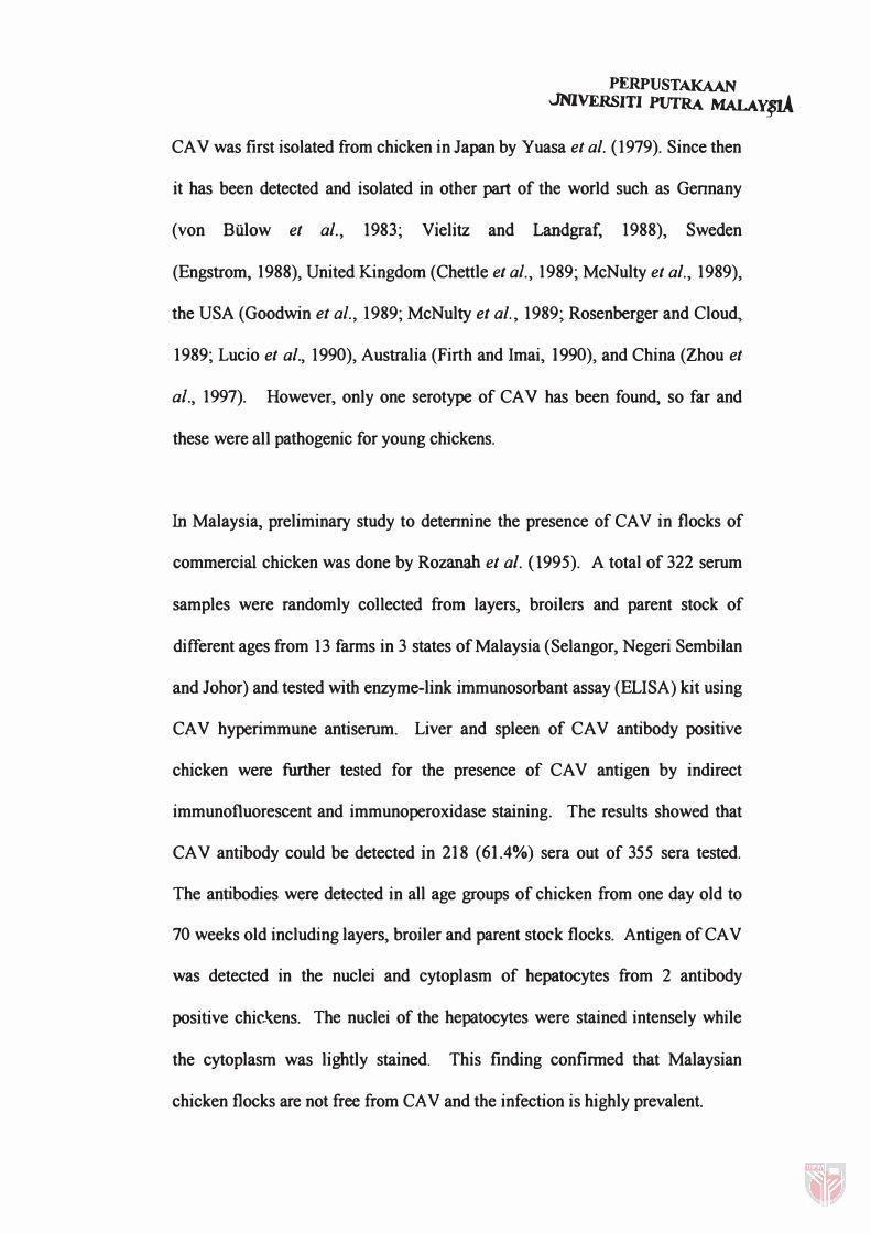
CAY was first isolated from chicken in Japan by Yuasa et al. (1979). Since then
it has been detected and isolated in other part of the world such as Gennany
(von Bulow et aI. , 1983; Vielitz and Landgraf, 1988), Sweden
(Engstrom, 1988), United Kingdom (Chettle et aI. , 1989; McNulty et al., 1989),
the USA (Goodwin et aI. , 1989; McNulty et al., 1989; Rosenberger and Cloud,
1989; Lucio et al., 1990), Australia (Firth and Imai, 1990), and China (Zhou et
af., 1997). However, only one serotype of CAY has been found, so far and
these were all pathogenic for young chickens.
In Malaysia, preliminary study to detennine the presence of CA V in flocks of
commercial chicken was done by Rozanah et af. (1 995). A total of 322 serum
samples were randomly collected from layers, broilers and parent stock of
different ages from 13 farms in 3 states of Malaysia (Selangor, Negeri Sembi Ian
and Johor) and tested with enzyme-link immunosorbant assay (ELISA) kit using
CA V hyperimmune antiserum. Liver and spleen of CA V antibody positive
chicken were further tested for the presence of CA V antigen by indirect
immunofluorescent and immunoperoxidase staining. The results showed that
CAY antibody could be detected in 218 (61.4%) sera out of 355 sera tested.
The antibodies were detected in all age groups of chicken from one day old to
70 weeks old including layers, broiler and parent stock flocks. Antigen of CA V
was detected in the nuclei and cytoplasm of hepatocytes from 2 antibody
positive chickens. The nuclei of the hepatocytes were stained intensely while
the cytoplasm was lightly stained. This finding confinned that Malaysian
chicken flocks are not free from CA V and the infection is highly prevalent.
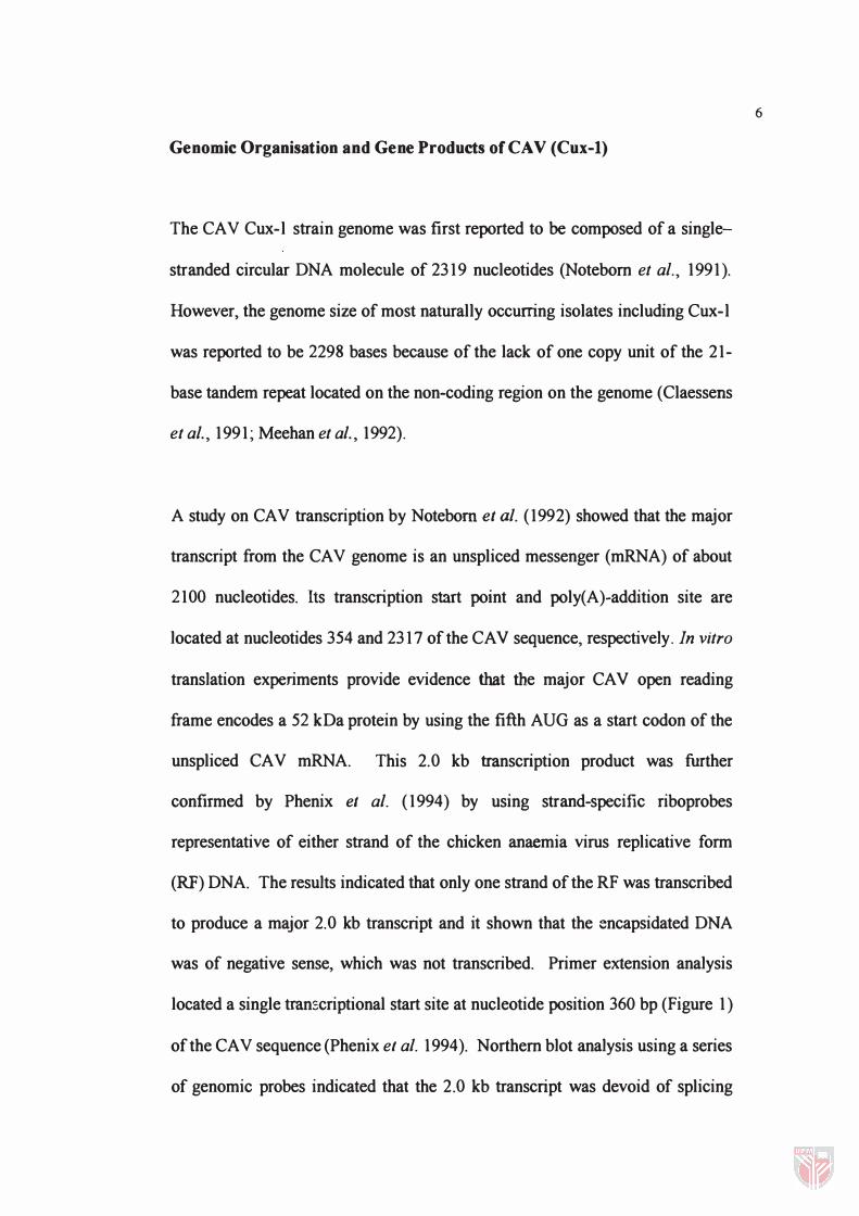
Genomic Organisation and Gene Products of CAV (Cux-I)
The CA V Cux-l strain genome was first reported to be composed of a single
stranded circular DNA molecule of 2319 nucleotides (Noteborn et aI. , 1991).
However, the genome size of most naturally occurring isolates including Cux-l
was reported to be 2298 bases because of the lack of one copy unit of the 21-
base tandem repeat located on the non-coding region on the genome (Claessens
et al. , 1991; Meehan et al. , 1992).
A study on CAY transcription by Notebom et al. (1992) showed that the major
transcript from the CA V genome is an unspliced messenger (mRNA) of about
2100 nucleotides. Its transcription start point and poly(A)-addition site are
located at nucleotides 354 and 2317 of the CA V sequence, respectively. In vitro
translation experiments provide evidence that the major CA V open reading
frame encodes a 52 kDa protein by using the fifth AUG as a start codon of the
unspliced CAY mRNA. This 2.0 kb transcription product was further
confirmed by Phenix el al. (1994) by using strand-specific riboprobes
representative of either strand of the chicken anaemia virus replicative form
(RF) DNA. The results indicated that only one strand of the RF was transcribed
to produce a major 2.0 kb transcript and it shown that the :;:ncapsidated DNA
was of negative sense, which was not transcribed. Primer extension analysis
located a single transcriptional start site at nucleotide position 360 bp (Figure 1)
of the CA V sequence (Phenix el al. 1994). Northern blot analysis using a series
of genomic probes indicated that the 2. 0 kb transcript was devoid of splicing
6
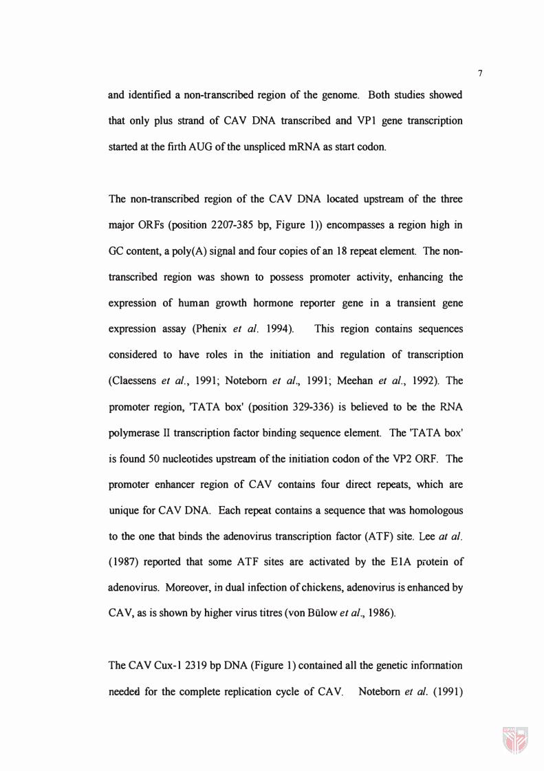
and identified a non-transcribed region of the genome. Both studies showed
that only plus strand of CA V DNA transcribed and VP1 gene transcription
started at the firth AUG of the unspliced mRNA as start codon.
The non-transcribed region of the CA V DNA located upstream of the three
major ORFs (position 2207-385 bp, Figure 1)) encompasses a region high in
GC content, a poly(A) signal and four copies of an 18 repeat element. The non
transcribed region was shown to possess promoter activity, enhancing the
expreSSIOn of human growth hormone reporter gene in a transient gene
expression assay (Phenix et a/. 1994). This region contains sequences
considered to have roles in the initiation and regulation of transcription
(Claessens et a/. , 1991; Notebom et a/., 1991; Meehan et aI. , 1992). The
promoter region, 'T AT A box' (position 329-336) is believed to be the RNA
polymerase II transcription factor binding sequence element. The 'T A T A box'
is found 50 nucleotides upstream of the initiation codon of the VP2 ORF. The
promoter enhancer region of CA V contains four direct repeats, which are
unique for CA V DNA. Each repeat contains a sequence that was homologous
to the one that binds the adenovirus transcription factor (ATF) site. Lee at a/.
(1987) reported that some ATF sites are activated by the E1A protein of
adenovirus. Moreover, in dual infection of chickens, adenovirus is enhanced by
CAY, as is shown by higher virus titres (von Bulow et a/., 1986).
The CA V Cux-l 2319 bp DNA (Figure 1) contained all the genetic infonnation
needed for the complete replication cycle of CAY. Notebom et a/. (1991)
7


![Azah Aziz [Sumber Elektronik].pdf](https://static.fdokumen.site/doc/165x107/587a19821a28abdd208b9a62/azah-aziz-sumber-elektronikpdf.jpg)