


Bahasa
Halaman
Undang-undang


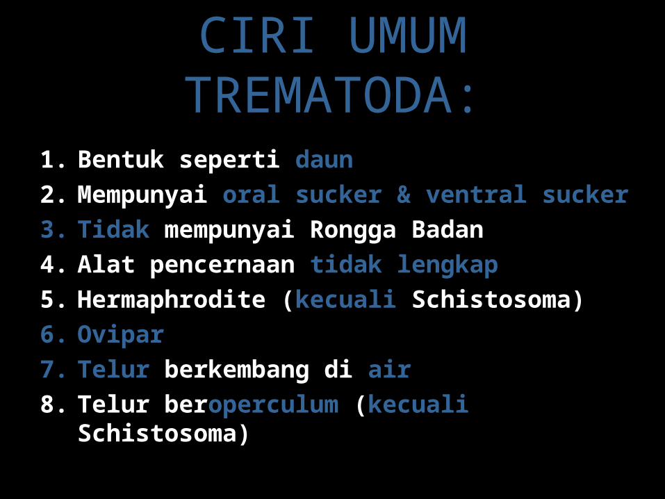
CIRI UMUM TREMATODA:
1. Bentuk seperti daun2. Mempunyai oral sucker & ventral sucker3. Tidak mempunyai Rongga Badan 4. Alat pencernaan tidak lengkap5. Hermaphrodite (kecuali Schistosoma)6. Ovipar7. Telur berkembang di air8. Telur beroperculum (kecuali Schistosoma)
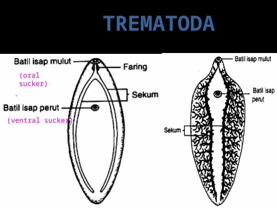
(oral sucker)
Ventral sucker
TREMATODA
(ventral sucker)
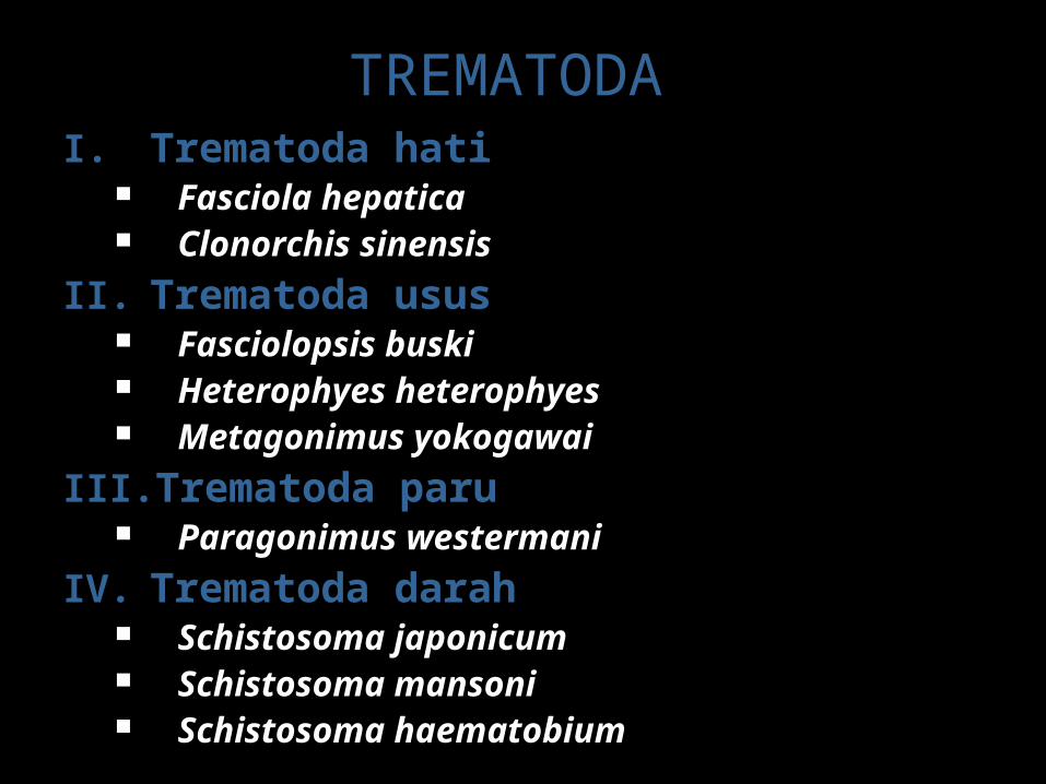
I. Trematoda hati Fasciola hepatica Clonorchis sinensis
II. Trematoda usus Fasciolopsis buski Heterophyes heterophyes Metagonimus yokogawai
III. Trematoda paru Paragonimus westermani
IV. Trematoda darah Schistosoma japonicum Schistosoma mansoni Schistosoma haematobium
TREMATODA

Fasciolopsis buski
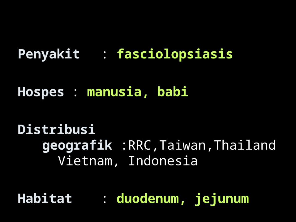
Penyakit: fasciolopsiasis
Hospes : manusia, babi
Distribusi geografik :RRC,Taiwan,Thailand Vietnam, Indonesia
Habitat : duodenum, jejunum
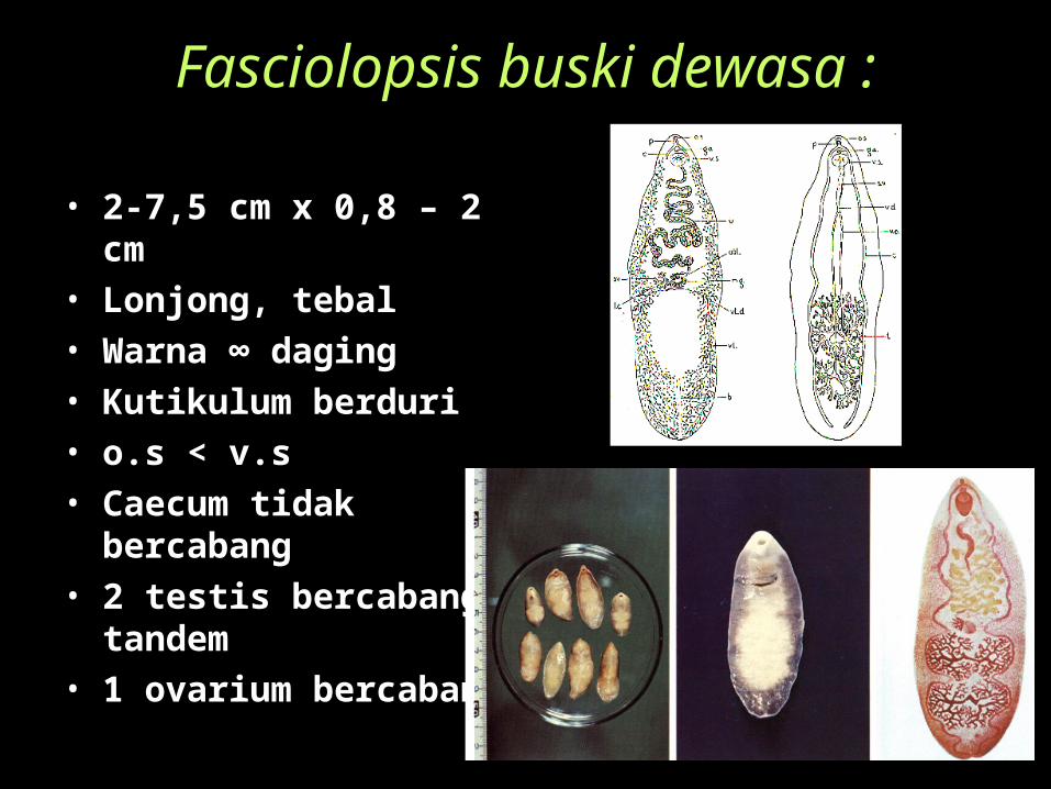
Fasciolopsis buski dewasa :
• 2-7,5 cm x 0,8 – 2 cm• Lonjong, tebal• Warna ∞ daging• Kutikulum berduri• o.s < v.s• Caecum tidak
bercabang• 2 testis bercabang,
tandem• 1 ovarium bercabang

TELUR Fasciolopsis buski
• 130-140 x 80 – 85 µ• Lonjong• Kekuningan• Operkulum • Dinding tipis, jernih
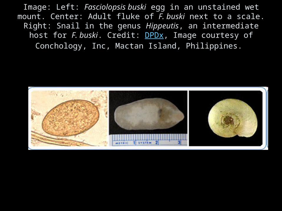
Image: Left: Fasciolopsis buski egg in an unstained wet mount. Center: Adult fluke of F. buski next to a scale. Right: Snail in the genus
Hippeutis, an intermediate host for F. buski. Credit: DPDx, Image courtesy of Conchology, Inc, Mactan Island, Philippines.
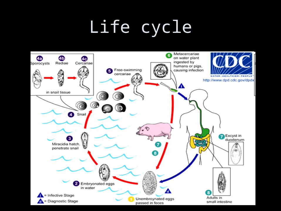
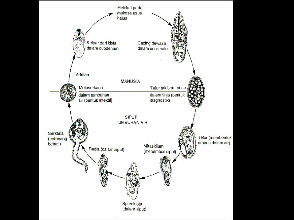
Life cycle
Life cycle• immature eggs are discharged into the intestine and stool . • Eggs become embryonated in water , eggs release miracidia ,
which invade a suitable snail intermediate host . • In the snail the parasites undergo several developmental stages
(sporocysts , rediae , and cercariae ). • The cercariae are released from the snail and encyst as
metacercariae on aquatic plants . • The mammalian hosts become infected by ingesting metacercariae
on the aquatic plants. After ingestion, the metacercariae excyst in the duodenum and attach to the intestinal wall.
• There they develop into adult flukes (20 to 75 mm by 8 to 20 mm) in approximately 3 months, attached to the intestinal wall of the mammalian hosts (humans and pigs) .
• The adults have a life span of about one year.
Gejala : nyeri epigastrium mual diare edema ileus akut
Diagnosa :gx klinis
(bila didaerah endemi)menemukan telur dlm tinja
Terapi:• diklorofen• niklosamid• praziquantel

Pencegahan• Memasak atau merendam
dalam air mendidih tumbuhan air yang akan dimakan
Heterophyes heterophyes( Penyakit :Heterophyasis )
• Distribusi geografis- Mesir- Palestina- Jepang- Cina- Korea- Taiwan- Filipina- Indonesia
• HabitatMukosa usus
• Definitif hostmanusia , mamalia
• Intermediate hostI. Pironella conica Cerithidea cingulataII. Tilapia nilotica Mugil japonicus Acanthogobius
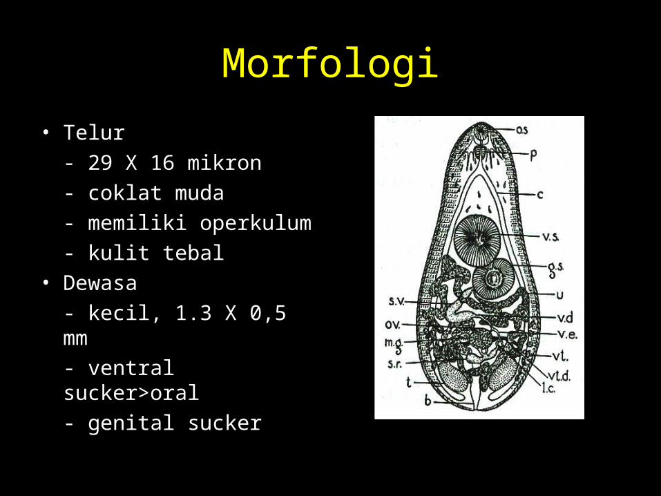
Morfologi• Telur
- 29 X 16 mikron- coklat muda- memiliki operkulum- kulit tebal
• Dewasa- kecil, 1.3 X 0,5 mm- ventral sucker>oral- genital sucker
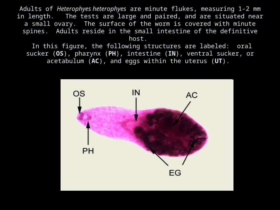
Adults of Heterophyes heterophyes are minute flukes, measuring 1-2 mm in length. The tests are large and paired, and are situated near a small ovary. The surface of the worm is covered with minute spines. Adults reside in the small intestine of the definitive host.
In this figure, the following structures are labeled: oral sucker (OS), pharynx (PH), intestine (IN), ventral sucker, or acetabulum (AC), and eggs within the uterus (UT).
Life cycle
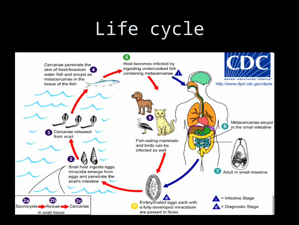
Life cycle• Adults release embryonated eggs each with a fully-developed
miracidium, and eggs are passed in the host's feces . After ingestion by a suitable snail (first intermediate host), the eggs hatch and release miracidia which penetrate the snail’s intestine . Genera Cerithidia and Pironella are important snail hosts in Asia and the Middle East respectively. The miracidia undergo several developmental stages in the snail, i.e. sporocysts , rediae , and cercariae . Many cercariae are produced from each redia. The cercariae are released from the snail and encyst as metacercariae in the tissues of a suitable fresh/brackish water fish (second intermediate host) . The definitive host becomes infected by ingesting undercooked or salted fish containing metacercariae . After ingestion, the metacercariae excyst, attach to the mucosa of the small intestine and mature into adults (measuring 1.0 to 1.7 mm by 0.3 to 0.4 mm) . In addition to humans, various fish-eating mammals (e.g., cats and dogs) and birds can be infected by Heterophyes heterophyes .

Gejala klinis
• Infeksi ringan- tidak tampak
• Infeksi berat- diare menahun- kolik- rasa tidak enak dan nyeri diperut- terdapat eosinofili
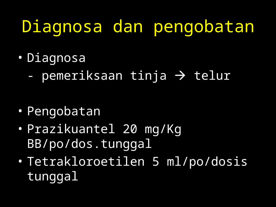
Diagnosa dan pengobatan
• Diagnosa- pemeriksaan tinja telur
• Pengobatan• Prazikuantel 20 mg/Kg BB/po/dos.tunggal• Tetrakloroetilen 5 ml/po/dosis tunggal
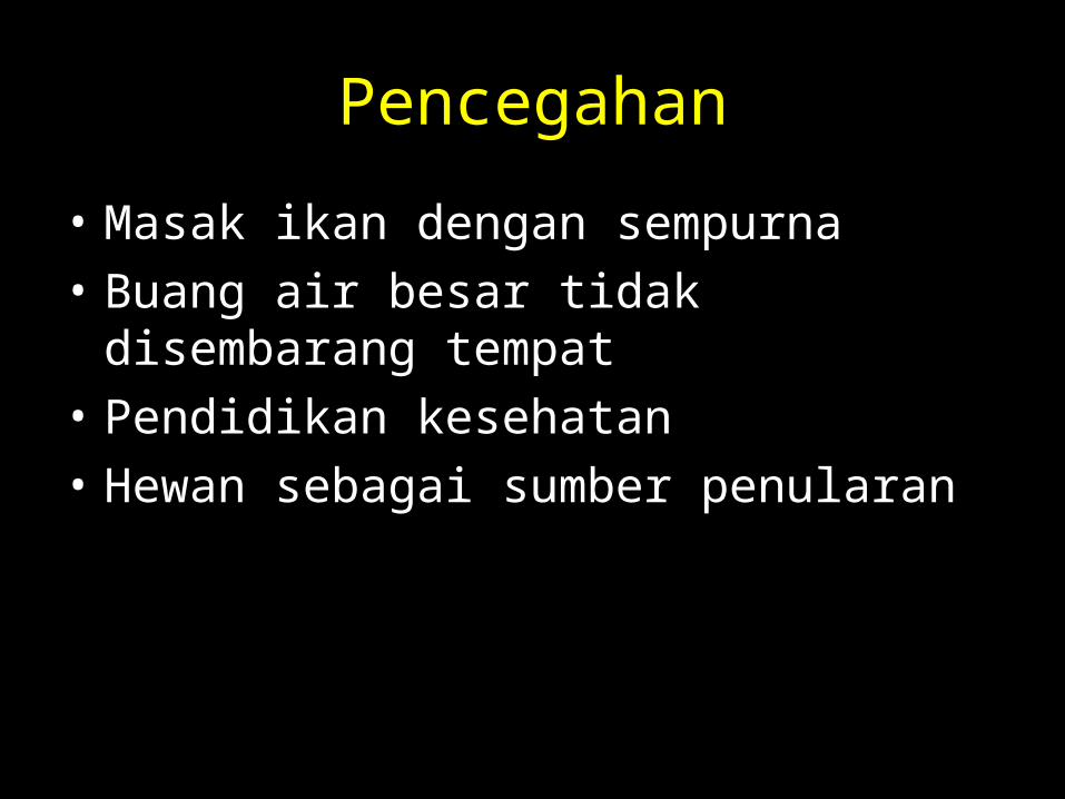
Pencegahan
• Masak ikan dengan sempurna• Buang air besar tidak disembarang tempat• Pendidikan kesehatan• Hewan sebagai sumber penularan
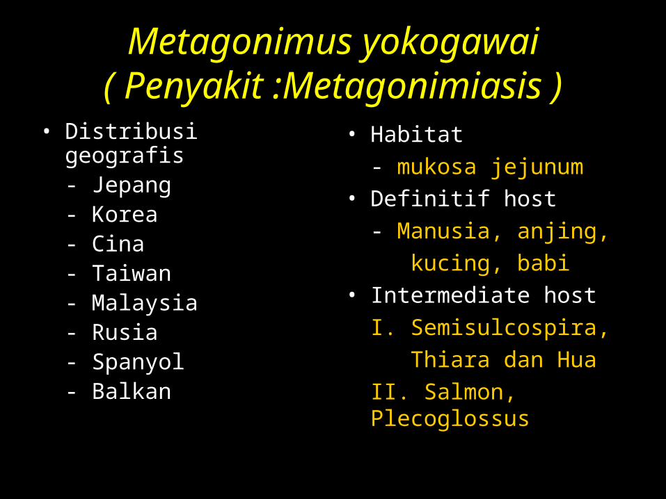
Metagonimus yokogawai( Penyakit :Metagonimiasis )
• Distribusi geografis- Jepang- Korea- Cina- Taiwan- Malaysia- Rusia- Spanyol- Balkan
• Habitat- mukosa jejunum
• Definitif host- Manusia, anjing, kucing, babi
• Intermediate hostI. Semisulcospira, Thiara dan HuaII. Salmon, Plecoglossus
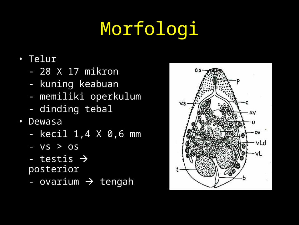
Morfologi• Telur
- 28 X 17 mikron- kuning keabuan- memiliki operkulum- dinding tebal
• Dewasa- kecil 1,4 X 0,6 mm- vs > os- testis posterior- ovarium tengah
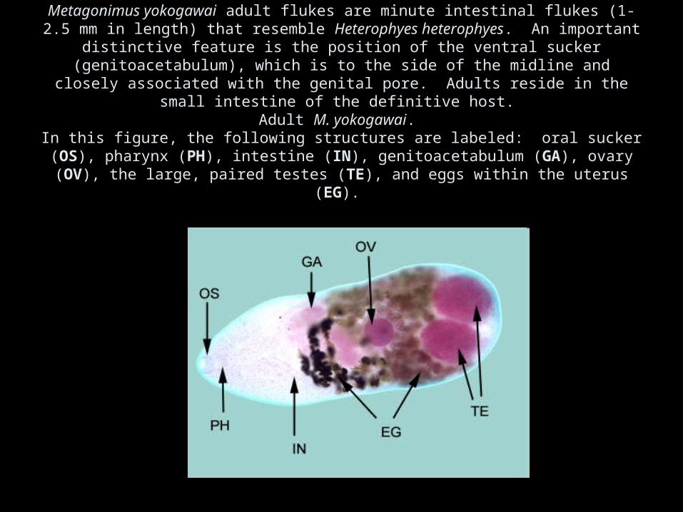
Metagonimus yokogawai adult flukes are minute intestinal flukes (1-2.5 mm in length) that resemble Heterophyes heterophyes. An important distinctive feature is the position of the
ventral sucker (genitoacetabulum), which is to the side of the midline and closely associated with the genital pore. Adults reside in the small intestine of the definitive host.
Adult M. yokogawai. In this figure, the following structures are labeled: oral sucker (OS), pharynx (PH),
intestine (IN), genitoacetabulum (GA), ovary (OV), the large, paired testes (TE), and eggs within the uterus (EG).
Life cycle
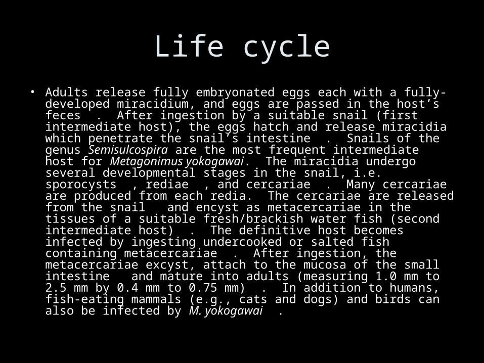
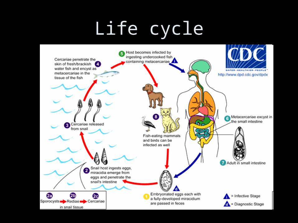
Life cycle• Adults release fully embryonated eggs each with a fully-developed
miracidium, and eggs are passed in the host’s feces . After ingestion by a suitable snail (first intermediate host), the eggs hatch and release miracidia which penetrate the snail’s intestine . Snails of the genus Semisulcospira are the most frequent intermediate host for Metagonimus yokogawai. The miracidia undergo several developmental stages in the snail, i.e. sporocysts , rediae , and cercariae . Many cercariae are produced from each redia. The cercariae are released from the snail and encyst as metacercariae in the tissues of a suitable fresh/brackish water fish (second intermediate host) . The definitive host becomes infected by ingesting undercooked or salted fish containing metacercariae . After ingestion, the metacercariae excyst, attach to the mucosa of the small intestine and mature into adults (measuring 1.0 mm to 2.5 mm by 0.4 mm to 0.75 mm) . In addition to humans, fish-eating mammals (e.g., cats and dogs) and birds can also be infected by M. yokogawai .

Gejala klinis
• Infeksi ringan tanpa gejala• Infeksi sedang diare dan sakit perut• Infeksi berat
- demam- nyeri perut- kolik- eosinofili
Diagnosa dan pengobatan
• Diagnosa- menemukan telur dalam tinja
• Pengobatan- Prazikuantel 20 mg/Kg BB/po/tunggal- Tetrakloroetilen 5 ml/po/tunggal
Pencegahan
• Masak ikan dengan sempurna• BAB tidak disembarang tempat• Pengawasan Hewan sumber penularan• Hygiene pribadi
Top Related