3rd e-Newsletter CTiG
4
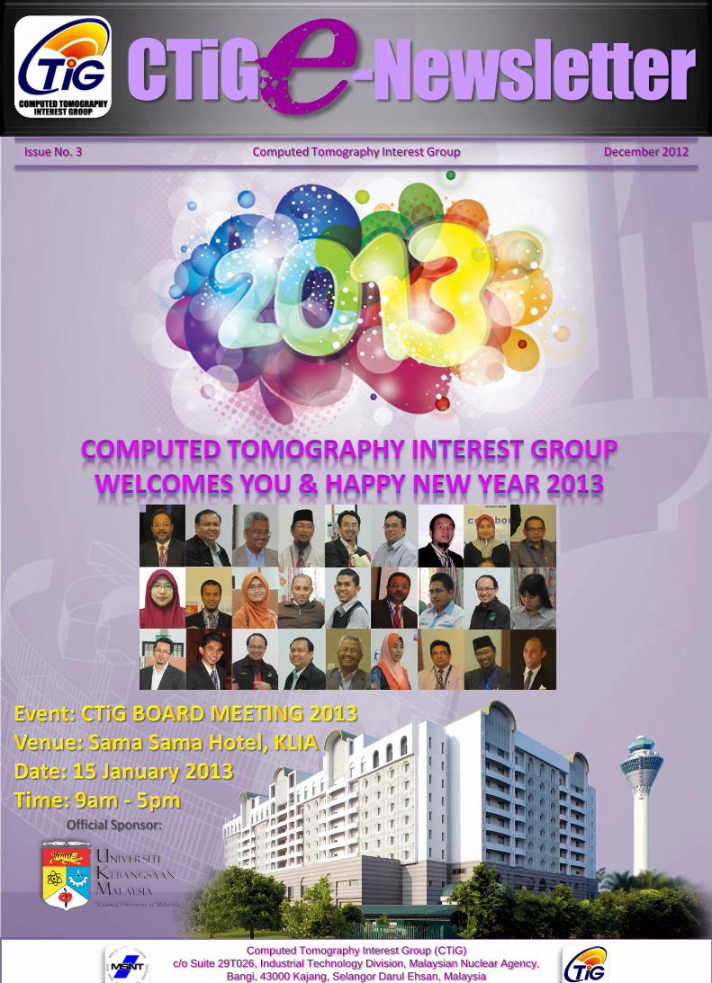
CTiG -Newsletter e Computed Tomography Interest Group (CTiG) c/o Suite 29T026, Industrial Technology Division, Malaysian Nuclear Agency, Bangi, 43000 Kajang, Selangor Darul Ehsan, Malaysia Tel: 03-89250510 Fax: 03-89250907 E-mail: [email protected] Issue No. 3 Computed Tomography Interest Group December 2012 Official Sponsor:
-
Upload
engku-fahmi -
Category
News & Politics
-
view
192 -
download
0
Transcript of 3rd e-Newsletter CTiG
CTiG -Newslettere
Computed Tomography Interest Group (CTiG)
c/o Suite 29T026, Industrial Technology Division, Malaysian Nuclear Agency,
Bangi, 43000 Kajang, Selangor Darul Ehsan, Malaysia
Tel: 03-89250510 Fax: 03-89250907 E-mail: [email protected]
Issue No. 3 Computed Tomography Interest Group December 2012
Official Sponsor:
Computed Tomography Interest Group (CTiG)
c/o Suite 29T026, Industrial Technology Division, Malaysian Nuclear Agency,
Bangi, 43000 Kajang, Selangor Darul Ehsan, Malaysia
Tel: 03-89250510 Fax: 03-89250907 E-mail: [email protected]
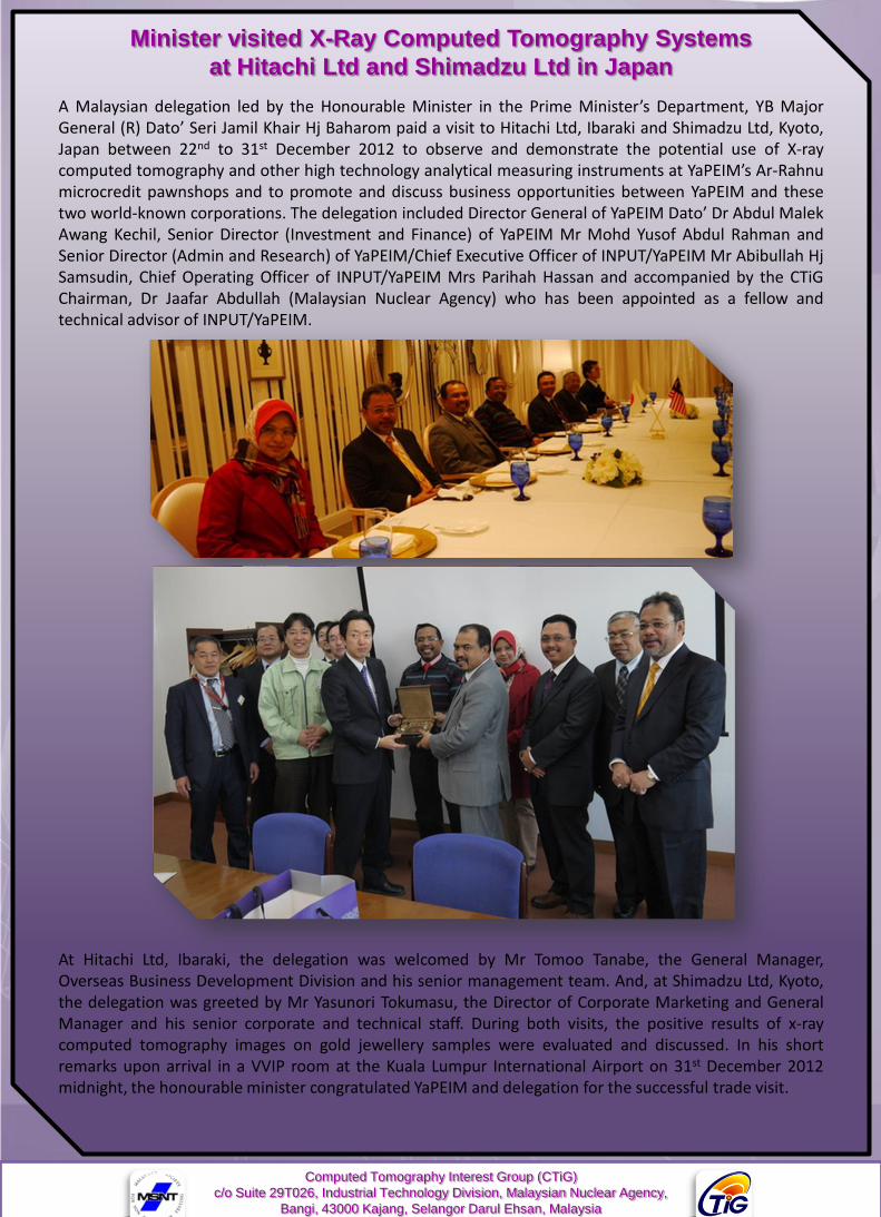
A Malaysian delegation led by the Honourable Minister in the Prime Minister’s Department, YB MajorGeneral (R) Dato’ Seri Jamil Khair Hj Baharom paid a visit to Hitachi Ltd, Ibaraki and Shimadzu Ltd, Kyoto,Japan between 22nd to 31st December 2012 to observe and demonstrate the potential use of X-raycomputed tomography and other high technology analytical measuring instruments at YaPEIM’s Ar-Rahnumicrocredit pawnshops and to promote and discuss business opportunities between YaPEIM and thesetwo world-known corporations. The delegation included Director General of YaPEIM Dato’ Dr Abdul MalekAwang Kechil, Senior Director (Investment and Finance) of YaPEIM Mr Mohd Yusof Abdul Rahman andSenior Director (Admin and Research) of YaPEIM/Chief Executive Officer of INPUT/YaPEIM Mr Abibullah HjSamsudin, Chief Operating Officer of INPUT/YaPEIM Mrs Parihah Hassan and accompanied by the CTiGChairman, Dr Jaafar Abdullah (Malaysian Nuclear Agency) who has been appointed as a fellow andtechnical advisor of INPUT/YaPEIM.
At Hitachi Ltd, Ibaraki, the delegation was welcomed by Mr Tomoo Tanabe, the General Manager,Overseas Business Development Division and his senior management team. And, at Shimadzu Ltd, Kyoto,the delegation was greeted by Mr Yasunori Tokumasu, the Director of Corporate Marketing and GeneralManager and his senior corporate and technical staff. During both visits, the positive results of x-raycomputed tomography images on gold jewellery samples were evaluated and discussed. In his shortremarks upon arrival in a VVIP room at the Kuala Lumpur International Airport on 31st December 2012midnight, the honourable minister congratulated YaPEIM and delegation for the successful trade visit.
Minister visited X-Ray Computed Tomography Systems
at Hitachi Ltd and Shimadzu Ltd in Japan
Cone Beam Computed Tomography (CBCT)By: Maya Genisa, PhD Candidate, School of Dental Sciences, USM.
CBCT is cone-beam scanners based on a cone-shaped beam of x-rays rotating around the object of interest
giving a volume of data, using a 2-dimensional extended digital array as an area detector. The technique involves
a single 360 degree scan in which the x-ray source and reciprocating area detector synchronously move around
the patient’s head, which is stabilized with a head holder.
CBCT image gathering consist of two phases; the first phase is data acquisition that uses cone shape x-ray beam
and the second phase is image reconstruction from computed tomography. The three dimensional images
(sagittal, coronal and axial plane) are produced using back projection inversion technique.
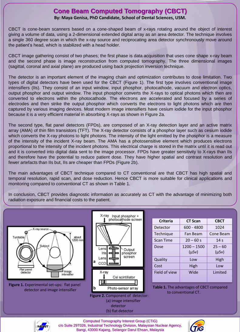
The detector is an important element of the imaging chain and optimization contributes to dose limitation. Two
types of digital detectors have been used for the CBCT (Figure 1). The first type involves conventional image
intensifiers (IIs). They consist of an input window, input phosphor, photocathode, vacuum and electron optics,
output phosphor and output window. The input phosphor converts the X-rays to optical photons which then are
converted to electrons within the photocathode. The electrons are accelerated and focused by a series of
electrodes and then strike the output phosphor which converts the electrons to light photons which are then
captured by various imaging devices. Most modern image intensifiers have cesium iodide for the input phosphor
because it is a very efficient material in absorbing X-rays as shown in Figure 2a.
The second type, flat panel detectors (FPDs), are composed of an X-ray detection layer and an active matrix
array (AMA) of thin film transistors (TFT). The X-ray detector consists of a phosphor layer such as cesium iodide
which converts the X-ray photons to light photons. The intensity of the light emitted by the phosphor is a measure
of the intensity of the incident X-ray beam. The AMA has a photosensitive element which produces electrons
proportional to the intensity of the incident photons. This electrical charge is stored in the matrix until it is read out
and it is converted into digital data sent to the image processor. FPDs have greater sensitivity to X-rays than IIs
and therefore have the potential to reduce patient dose. They have higher spatial and contrast resolution and
fewer artefacts than IIs but, IIs are cheaper than FPDs (Figure 2b).
The main advantages of CBCT technique compared to CT conventional are that CBCT has high spatial and
temporal resolution, rapid scan, and dose reduction. Hence CBCT is more suitable for clinical applications and
monitoring compared to conventional CT as shown in Table 1.
In conclusion, CBCT provides diagnostic information as accurately as CT with the advantage of minimizing both
radiation exposure and financial costs to the patient.
Computed Tomography Interest Group (CTiG)
c/o Suite 29T026, Industrial Technology Division, Malaysian Nuclear Agency,
Bangi, 43000 Kajang, Selangor Darul Ehsan, Malaysia
Tel: 03-89250510 Fax: 03-89250907 E-mail: [email protected]
Criteria CT Scan CBCT
Detector 600 - 4800 1024
Technique Fan Beam Cone Beam
Scan Time 20 – 60 s 14 s
Dose 1200 – 1500 (µSv)
25 – 60 (µSv)
Quality Low High
Cost High Low
Field of view Wide Limited
a
b Table 1. The advantages of CBCT comparedto conventional CT.
Figure 2. Component of detector: (a) image intensifier
detector (b) flat detector
Figure 1. Experimental set-ups: flat panel detector and image intensifier
From the Editorial Desk….
Chief Editor
Assoc. Prof. Dr. Zainul Ahmad Rajion
Dear Colleagues,
First of all, on behalf of the CTiG editorial group, I would liketo wish all CTiG members a happy new year 2013. Recently,with the overwhelming responses in number of CTiGmemberships, it is important for us to strengthen ourpotential collaboration. For a start, we can have jointresearch or joint supervision/co-supervision amonginstitutions and universities to fully utilize our resources. AllCTiG members are encouraged to get involved with inter-disciplinary research projects by communicating, sharingideas and collaborating across disciplines.
As a new year has already started, why don’t we set one ofour intentions to publish an article in the CTiG newsletter? Bypublishing our works, other researchers can understandmore about our research. On a personal level, it is verysatisfying to see the results of your hard work going into printand being read by academics and researchers. Not only forthe recognition of our efforts and self-satisfaction, it can alsoconsolidate our knowledge and even lead to new discovery.In other words, publishing is an integral part of the discoveryof new ideas. Therefore, I hope that we can see more articlesfrom other members in the next newsletter.
Board Members of CTiG 2012/2013
Chairman : Dr. Jaafar Abdullah (ANM)Deputy Chairman : Prof. Dr. Ruzairi Abdul Rahim (UTM)Hon. Secretary : Salzali Mohd (ANM)Deputy Hon. Secretary : Engku Mohd Fahmi Engku Chik(ANM) Hon. Treasurer : Dr. Elmy Johana Mohamad (UTHM)
Committee Members :•Prof. Ir. Dr. Mohd Sobri Takriff (UKM)•Assoc. Prof Dr. Zainul Ahmad Rajion (USM)•Assoc. Prof. Ir. Dr. Idris Ismail (UTP)•Assoc. Prof. Dr. Sam’an Malik Masudi (USM)•Mohd Hafiz Fazalul Rahiman (UniMAP)•Dr. Mohd Hezri Fazalul Rahiman (UiTM)•Yasmin Abdul Wahab (UMP)•Suzanna Ridzuan Aw (TATIUC)•Mohd Amirul Syafiq Mohd Yunos (ANM)•Roslan Yahya (ANM)
CTiG e-Newsletter is in the process of collectingrelated articles on Computed Tomography. Theacademics, professionals and researchers arewelcome to contribute to the success of the e-Newsletter since increase growth in the number ofCTiG memberships since 2011.
Please contact the Chief Editor, Assoc. Prof. Dr. Zainul Ahmad Rajion at [email protected] for submission.
Computed Tomography Interest Group (CTiG)
c/o Suite 29T026, Industrial Technology Division, Malaysian Nuclear Agency,
Bangi, 43000 Kajang, Selangor Darul Ehsan, Malaysia
Tel: 03-89250510 Fax: 03-89250907 E-mail: [email protected]
IAEA/RCA Regional Training Course on
Monte Carlo Simulations for RPT, CT,
SPECT and Design of Radiotracer
Experiments in Daejeon, Korea
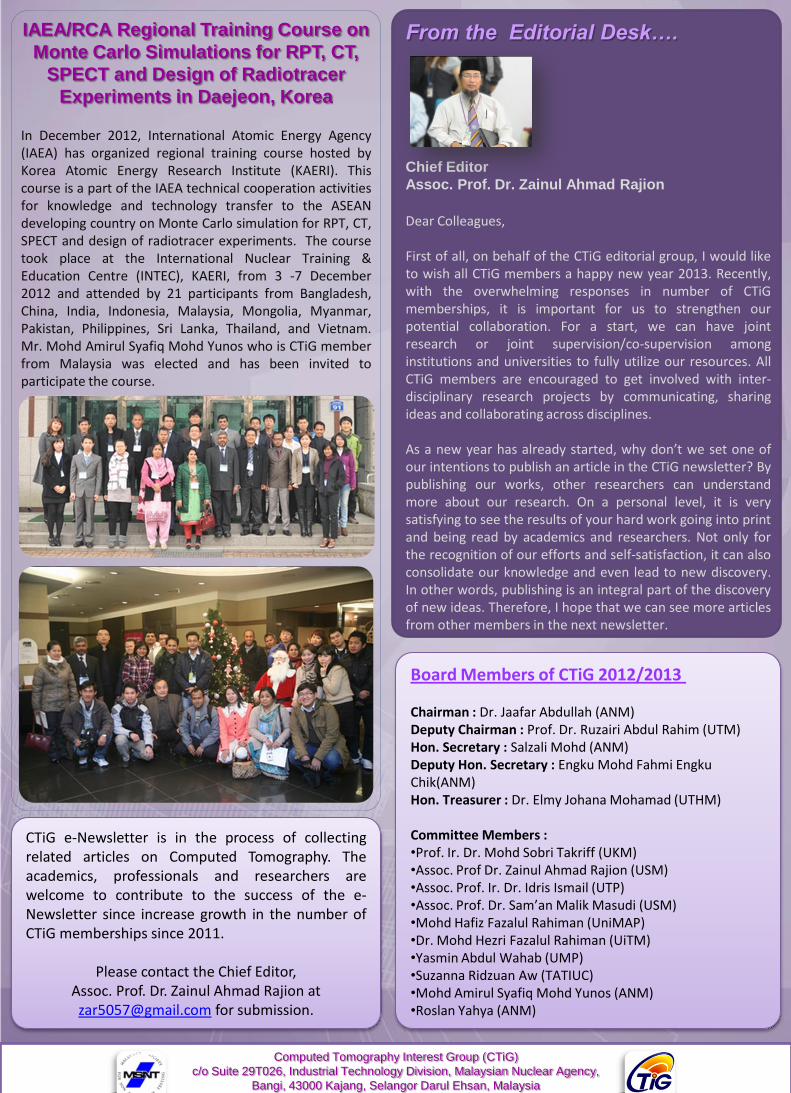
In December 2012, International Atomic Energy Agency(IAEA) has organized regional training course hosted byKorea Atomic Energy Research Institute (KAERI). Thiscourse is a part of the IAEA technical cooperation activitiesfor knowledge and technology transfer to the ASEANdeveloping country on Monte Carlo simulation for RPT, CT,SPECT and design of radiotracer experiments. The coursetook place at the International Nuclear Training &Education Centre (INTEC), KAERI, from 3 -7 December2012 and attended by 21 participants from Bangladesh,China, India, Indonesia, Malaysia, Mongolia, Myanmar,Pakistan, Philippines, Sri Lanka, Thailand, and Vietnam.Mr. Mohd Amirul Syafiq Mohd Yunos who is CTiG memberfrom Malaysia was elected and has been invited toparticipate the course.