First Trimester Prenatal Diagnosis of Beta-thalassaemia following … · NYK was a 32 year old...
4
CASE REPORT First Trimester Prenatal Diagnosis of Beta-thalassaemia following Chorionic Villus Sampling R. Chandran, MRCOG* O. Ainoon, DCP** I. Anson, DipMLT* J. Anne, DipMLT** S.K. Cheong, FRCPA** * Department of Obstetrics and Gynaecology, Universiti Kebangsaan Malaysia, Jalan Raia Muda Abdul Aziz, 50300 Kuala Lumpur ** Department of Pathology, Universiti Kebangsaan Malaysia, Jalan Raia Muda Abdul Aziz, 50300 Kuala Lumpur Introduction Beta-thalassaemia is a common condition in this country, afflicting mainly'the Chinese and Malays. Until recently, prenatal diagnosis of this condition was not available locally. We report here our preliminary experience of prenatal diagnosis of this condition in the first trimester following chorionic villus sampling( CVS). Case Reports Case 1 NYK was a 32 year old Chinese secundigravida who was referred at 10 weeks' gestation for prenatal diagnosis. Her first-born had beta-thalassaemia major and was transfusion depend- ent. Subsequent DNA analysis showed that both parents were heterozygous for the beta- thalassaemia mutation frameshift 41142 (-TCTT). An ultrasound scan using a Toshiba SSA-l OOA with a 3.75 mHz curvilinear transducer confirmed a viable intrauterine pregnancy corresponding to 10 weeks' gestation. The placental site was situated antero- laterally. After counselling, the parents consented to CVS. This was performed by one of us (RC) transabdominally under ultrasound guidance with a 20 gauge spinal needle using a free-hand technique. The needle tip was guided parallel to the chorionic plate and into the chorion frondosum. Villi were aspirated into heparinised buffered culture medium using a 10 ml syringe with a gentle to and fro movement of the sampling needle. An adequate sample was obtained at the first attempt. Anti-D immunoglobulin was not required as the patient was Rhesus positive. The parents were reassured and Med J Malaysia Vol 48 No 3 Sept 1993 341
-
Upload
nguyencong -
Category
Documents
-
view
226 -
download
0
Transcript of First Trimester Prenatal Diagnosis of Beta-thalassaemia following … · NYK was a 32 year old...
CASE REPORT
First Trimester Prenatal Diagnosis of Beta-thalassaemia following Chorionic Villus Sampling
R. Chandran, MRCOG* O. Ainoon, DCP**
I. Anson, DipMLT*
J. Anne, DipMLT**
S.K. Cheong, FRCPA** * Department of Obstetrics and Gynaecology, Universiti Kebangsaan Malaysia, Jalan Raia
Muda Abdul Aziz, 50300 Kuala Lumpur ** Department of Pathology, Universiti Kebangsaan Malaysia, Jalan Raia Muda Abdul Aziz,
50300 Kuala Lumpur
Introduction
Beta-thalassaemia is a common condition in this country, afflicting mainly'the Chinese and Malays. Until recently, prenatal diagnosis of this condition was not available locally. We report here our preliminary experience of prenatal diagnosis of this condition in the first trimester following chorionic villus sampling( CVS).
Case Reports
Case 1
NYK was a 32 year old Chinese secundigravida who was referred at 10 weeks' gestation for prenatal diagnosis. Her first-born had beta-thalassaemia major and was transfusion dependent. Subsequent DNA analysis showed that both parents were heterozygous for the betathalassaemia mutation frameshift 41142 (-TCTT).
An ultrasound scan using a Toshiba SSA-l OOA with a 3.75 mHz curvilinear transducer confirmed a viable intrauterine pregnancy corresponding to 10 weeks' gestation. The placental site was situated anterolaterally. After counselling, the parents consented to CVS. This was performed by one of us (RC) transabdominally under ultrasound guidance with a 20 gauge spinal needle using a free-hand technique. The needle tip was guided parallel to the chorionic plate and into the chorion frondosum. Villi were aspirated into heparinised buffered culture medium using a 10 ml syringe with a gentle to and fro movement of the sampling needle. An adequate sample was obtained at the first attempt. Anti-D immunoglobulin was not required as the patient was Rhesus positive. The parents were reassured and
Med J Malaysia Vol 48 No 3 Sept 1993 341
CASE REPORT
shown the fetal heart activity after the procedure. The patient was advised to avoid strenuous activity for a week and she did not have any procedure-related complications such as pain, fever or vaginal bleeding.
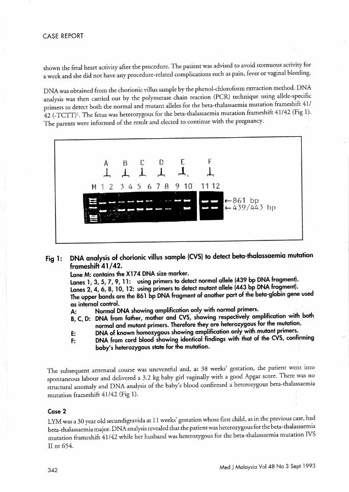
DNA was obtained from the chorionic villus sample by the phenol-chloroform extraction method. DNA analysis was then carried out by the polymerase chain reaction (PCR) technique using allele-specific primers to detect both the normal and mutant alleles for the beta-thalassaemia mutation frameshift 411 42 (-TCTT)l. The fetus was heterozygous for the beta-thalassaemia mutation frameshift 41/42 (Fig 1). The parents were informed of the result and elected to continue with the pregnancy.
A B C o E F 1, L J., L ,.1" l,
M 1 2 3 4 5 6 7 8 9 10' 1112
-::;=:..-.---- .... - ... -- ........
.---- .... "-,----- .... _--- -; t=:I) '=. ~
1iIf-861 bp ~439/443 bp
Fig 1: DNA analysis of chorionic villus sample (CVS) to detect beta-thalassaemia mutation frameshift 41/42. Lane M: contains the X174 DNA size marker .
. Lanes 1,3,5,7,9, 11: using primers to detect normal allele (439 bp DNA fragment). Lanes 2, 4, 6, 8, 10, 12: using primers to detect mutant allele (443 bp DNA fragment). The upper bands are the 861 bp DNA fragment of another part of the beta-globin gene us~d as internal control. A: Normal DNA showing amplification only with normal primers. B, C, D: DNA from father, mother and CVS, showing respectively amplification with. both
normal and mutant primers. Therefore they are heterozygous for the mutation, E: DNA of known homozygous showing amplification only with mutant primers .. F: DNA from cord blood showing identical findings with that of the CYS, confirming
baby's heterozygous state for the mutation.
<,
The subsequent antenatal course was uneventful and, at 38 weeks' gestation, the patient went into spontaneous labour and delivered a 3.2 kg baby girl vaginally with a good Apgar score. There was no structural anomaly and DNA analysis of the baby's blood confirmed a heterozygous beta-thalassaemia mutation frameshift 41142 (Fig 1).
Case 2
LYM was a 30 year old secundigravida at 11 weeks' gestation whose first child, as in the previous case, had beta-thalassaemiamajor. DNA analysis revealed that the patient was heterozygous for the beta-thalassaemia mutation frameshift 41142 while her husband was heterozygous for the beta-thalassaemia mutation IVS II nt 654.
342 Med J Malaysia Vol 48 No 3 Sept 1993
FIRST TRIMESTER PRENATAL DIAGNOSIS OF BETA-THALASSAEMIA
An ultrasound scan confirmed a viable intrauterine pregnancy of 11 weeks' gestation with a posterior placental site. Mter appropriate counselling and consent, a transabdominal CVS was carried out successfully as described previously. This patient too was Rhesus positive and did not require anti-D immunoglobulin.
DNA analysis of the chorionic villus sample showed the fetus to be heterozygous for the betathalassaemia mutation frameshift 41/42. As in the previous case, the parents elected to continue with the pregnancy. This pregnancy is ongoing and there has been no procedurerelated complication to date at 20 weeks' gestation.
Discussion
Prenatal detection of beta-thalassaemia is a complex task because many different mutations can give rise to defective beta-globin chain synthesis. The use of synthetic oligonucleotides to detect directly a point mutation in the DNA that produces beta-thalassaemia was first described in 198Y, but has hitherto not been available in this country. This direct technique of using radiolabelled oligonucleotide probes has been further refined and it is now possible to use non-radioactive direct gene amplification techniques which are simpler and quicker.
Although it is possible to obtain fetal DNA from an amniotic fluid sample, the chorionic villus gives a better yield. It has been estimated that a single chorionic villus gives the same yield of DNA as the cells in 10 ml of amniotic fluid3• The additional advantage of CVS is that it affords diagnosis in the first trimester. If an abnormal result is obtained, parents can then be given the option of a termination in the first trimester which is not only a simpler and safer procedure, but also psychologically advantageous compared to a second trimester termination of pregnancy.
The main drawback ofCVS is the fetal loss rate of2% to 4%4. This is due in part to the higher natural loss rate in the first trimester but is also related to the experience of the operator and the number of sampling attempts. We have shown that it is possible to obtain an adequate sample for DNA analysis with the transabdominal technique even with a posteriorly situated placental site without early fetal loss. The first case carried uneventfully to term, while the second case is ongoing at 20 weeks' gestation. Both these patients did not have any vaginal bleeding after the procedure and this is a known advantage of the transabdominal technique.
Given the frequency of be ta-thalassaemia in this country, we believe that our results represent a significant development in the prenatal diagnosis of this condition in Malaysia. In order for couples to gain maximally from this type of first trimester diagnosis, medical practitioners and health workers should be aware that early referral is crucial so that appropriate and careful counselling can be carried out, ideally, even before a subsequent pregnancy is embarked upon.
Acknowledgement
We would like to thank our Dean, Professor Dato' Or KhalidAbdul Kadir, who has been a constant source of support and encouragement. The techniques of DNA analysis were made possible by an IRP A project grant (03-07-03-03).
Med J Malaysia Vol 48 No 3 Sept 1993 343
FIRST TRIMESTER PRENATAL DIAGNOSIS OF BETA-THALASSAEMIA
1. Old JM, Varawalla NY, Weathera11 DJ. Rapid detection and prenatal diagnosis of beta-thalassaemia: studies in Indian and Cypriot popularions in the UK. Lancet 1990;553 : 834-7.
2. Piratsu M, Kar YW, Cao A et aL Prenatal diagnosis of betathalassaemia: detection of a single nucleotide mutation in DNA. New Engl J Med 1983;309 : 284-7.
344
3. Williamson R, Eskdale J, Coleman DV et aL Direct gene analysis of chorionic villli: a possible technique for Hrst trimester antenatal diagnosis ofhaemoglobinopathies. Lancet 1981;ii: 1125-7.
4. Jackson LG, Wapner RJ. Risks of chorionic villus sampling. Clin Obstet Gynaeco11987;1 : 513-31.
Med J Malaysia Vol 48 No 3 Sep! 1993