Isolation and Photophysical Properties of Di- and Tri ... Nabila... · Seidel et al. 2013; Xu et...
6
Sains Malaysiana 47(5)(2018): 903–906 http://dx.doi.org/10.17576/jsm-2018-4705-05 Isolation and Photophysical Properties of Di- and Tri-substituted Natural Anthraquinones from Malaysian Morinda citrifolia (Pengasingan dan Sifat Fotofizikal Antraquinon Semula Jadi Tertukarganti-Di dan Tri daripada Morinda citrifolia di Malaysia) NABILA ELYANA ADNAN, NUR ATIQAH MOHD NASUHA, ZANARIAH ABDULLAH, YEUN-MUN CHOO* & HAIRUL ANUAR TAJUDDIN ABSTRACT Five di- and tri-substituted natural anthraquinones, i.e. nordamnacanthal (1), damnacanthal (2), rubiadin (3), 1-methoxy- 2-methyl-3-hydroxyanthraquinone (4) and 1-hydroxy-3-methoxyanthraquinone (5) were subjected to photophysical studies. The results indicated that steric hindrance and intramolecular hydrogen bonding are important factors that affect absorption and emission spectral of these natural anthraquinones. Besides that, emission properties were significantly enhanced with formation of intramolecular hydrogen bonding in 1,3-dihydroxy-2-aldehyde tri-substituted anthraquinone 1. This gave rise to formation of two additional quasi aromatic rings extending the π-conjugation system in the anthraquinone structure. Keywords: Absorption spectral; anthraquinone; emission spectral; intramolecular hydrogen bonding; photophysical properties ABSTRAK Lima antraquinon semula jadi, iaitu nordamnacanthal (1), damnacanthal (2), rubiadin (3), 1-metoksi-2-metil-3- hidroksiantraquinon (4) dan 1-hidroksi-3-metoksiantraquinon (5) digunakan dalam kajian sifat fotofizikal. Keputusan kajian menunjukkan bahawa penghalang sterik dan ikatan hidrogen intramolekul adalah faktor penting yang mempengaruhi spektrum penyerapan dan pelepasan antraquinon semula jadi. Selain itu, sifat fotofizikal pemancaran antraquinon 1 tertukarganti-tri 1,3-dihidroksi-2-aldehid dipertingkatkan dengan pembentukan ikatan hidrogen intramolekul. Pembentukan ikatan hidrogen intramolekul ini membolehkan pembentukan dua gelang aromatik kuasi tambahan dan memanjangkan sistem konjugasi-π dalam struktur antraquinon. Kata kunci: Antraquinon; ikatan hidrogen intramolekul; sifat fotofizikal; spektra pelepasan spektra penyerapan INTRODUCTION Anthraquinones are a group of naturally occurring important compounds that can also be obtained synthetically (Madje et al. 2010; Puenner et al. 2013; Seidel et al. 2013; Xu et al. 2010). The economic significance of anthraquinones such as in manufacture of dyes and pigments have caused advanced studies to be conducted, which focused on optical spectroscopy and photochemistry properties of the compound (Gordon & Gregory 1983). In addition, anthraquinones are utilised as photosensitizers, chelating agents in analytical applications and medicinal and pharmacological agents (Diaz 1990). Although reviews on anthraquinone derivatives’ absorption and emission spectral properties have been published previously (Diaz 1990; Langdon- Jones & Pope 2014), these investigations focused on synthetic anthraquinones with unnatural substitution groups. Nevertheless, studies on optical spectroscopy of natural anthraquinones, which possess different types and positions of substitution groups compared to synthetic anthraquinones, have not been comprehensively reported yet. Therefore, this presented a vacuum in information on anthraquinones’ optical properties. As part of our on-going research (Nur Atiqah & Choo 2016; Yap et al. 2016, 2015), a series of natural anthraquinones from the roots of Morinda citrifolia have been isolated and these pure compounds were subjected to photophysical studies. Morinda citrifolia is known by a variety of common names such as mengkudu in Malaysia, noni in Hawaii, ba ji tian in China, cheese fruit in Australia, Indian mulberry in India, monkey dumpling and forbidden fruit in Barbados, yaw weed in Guyana, nhau in Vietnam, nono in Tahiti, and nonu in Samoa and Tonga. M. citrifolia has been used in traditional medicine to treat a wide range of ailments, which include diabetes, diarrhea, hypentension, malaria, asthma, cancer, high blood pressure and arthritis. It has been reported in previous study that the roots of M. citrifolia is a rich source of anthraquinone compounds. In addition to its medicinal values, anthraquinones is an important ingredient in dye and pigment industries. The investigation of Malaysia M. citrifolia chemical constituents lead to the isolation of five anthraquinones,
Transcript of Isolation and Photophysical Properties of Di- and Tri ... Nabila... · Seidel et al. 2013; Xu et...

Sains Malaysiana 47(5)(2018): 903–906 http://dx.doi.org/10.17576/jsm-2018-4705-05
Isolation and Photophysical Properties of Di- and Tri-substituted Natural Anthraquinones from Malaysian Morinda citrifolia
(Pengasingan dan Sifat Fotofizikal Antraquinon Semula Jadi Tertukarganti-Di dan Tri daripada Morinda citrifolia di Malaysia)
NABILA ELYANA ADNAN, NUR ATIQAH MOHD NASUHA, ZANARIAH ABDULLAH, YEUN-MUN CHOO* & HAIRUL ANUAR TAJUDDIN
ABSTRACT
Five di- and tri-substituted natural anthraquinones, i.e. nordamnacanthal (1), damnacanthal (2), rubiadin (3), 1-methoxy-2-methyl-3-hydroxyanthraquinone (4) and 1-hydroxy-3-methoxyanthraquinone (5) were subjected to photophysical studies. The results indicated that steric hindrance and intramolecular hydrogen bonding are important factors that affect absorption and emission spectral of these natural anthraquinones. Besides that, emission properties were significantly enhanced with formation of intramolecular hydrogen bonding in 1,3-dihydroxy-2-aldehyde tri-substituted anthraquinone 1. This gave rise to formation of two additional quasi aromatic rings extending the π-conjugation system in the anthraquinone structure.
Keywords: Absorption spectral; anthraquinone; emission spectral; intramolecular hydrogen bonding; photophysical properties
ABSTRAK
Lima antraquinon semula jadi, iaitu nordamnacanthal (1), damnacanthal (2), rubiadin (3), 1-metoksi-2-metil-3-hidroksiantraquinon (4) dan 1-hidroksi-3-metoksiantraquinon (5) digunakan dalam kajian sifat fotofizikal. Keputusan kajian menunjukkan bahawa penghalang sterik dan ikatan hidrogen intramolekul adalah faktor penting yang mempengaruhi spektrum penyerapan dan pelepasan antraquinon semula jadi. Selain itu, sifat fotofizikal pemancaran antraquinon 1 tertukarganti-tri 1,3-dihidroksi-2-aldehid dipertingkatkan dengan pembentukan ikatan hidrogen intramolekul. Pembentukan ikatan hidrogen intramolekul ini membolehkan pembentukan dua gelang aromatik kuasi tambahan dan memanjangkan sistem konjugasi-π dalam struktur antraquinon.
Kata kunci: Antraquinon; ikatan hidrogen intramolekul; sifat fotofizikal; spektra pelepasan spektra penyerapan
INTRODUCTION
Anthraquinones are a group of naturally occurring important compounds that can also be obtained synthetically (Madje et al. 2010; Puenner et al. 2013; Seidel et al. 2013; Xu et al. 2010). The economic significance of anthraquinones such as in manufacture of dyes and pigments have caused advanced studies to be conducted, which focused on optical spectroscopy and photochemistry properties of the compound (Gordon & Gregory 1983). In addition, anthraquinones are utilised as photosensitizers, chelating agents in analytical applications and medicinal and pharmacological agents (Diaz 1990). Although reviews on anthraquinone derivatives’ absorption and emission spectral properties have been published previously (Diaz 1990; Langdon-Jones & Pope 2014), these investigations focused on synthetic anthraquinones with unnatural substitution groups. Nevertheless, studies on optical spectroscopy of natural anthraquinones, which possess different types and positions of substitution groups compared to synthetic anthraquinones, have not been comprehensively reported
yet. Therefore, this presented a vacuum in information on anthraquinones’ optical properties. As part of our on-going research (Nur Atiqah & Choo 2016; Yap et al. 2016, 2015), a series of natural anthraquinones from the roots of Morinda citrifolia have been isolated and these pure compounds were subjected to photophysical studies. Morinda citrifolia is known by a variety of common names such as mengkudu in Malaysia, noni in Hawaii, ba ji tian in China, cheese fruit in Australia, Indian mulberry in India, monkey dumpling and forbidden fruit in Barbados, yaw weed in Guyana, nhau in Vietnam, nono in Tahiti, and nonu in Samoa and Tonga. M. citrifolia has been used in traditional medicine to treat a wide range of ailments, which include diabetes, diarrhea, hypentension, malaria, asthma, cancer, high blood pressure and arthritis. It has been reported in previous study that the roots of M. citrifolia is a rich source of anthraquinone compounds. In addition to its medicinal values, anthraquinones is an important ingredient in dye and pigment industries. The investigation of Malaysia M. citrifolia chemical constituents lead to the isolation of five anthraquinones,
904
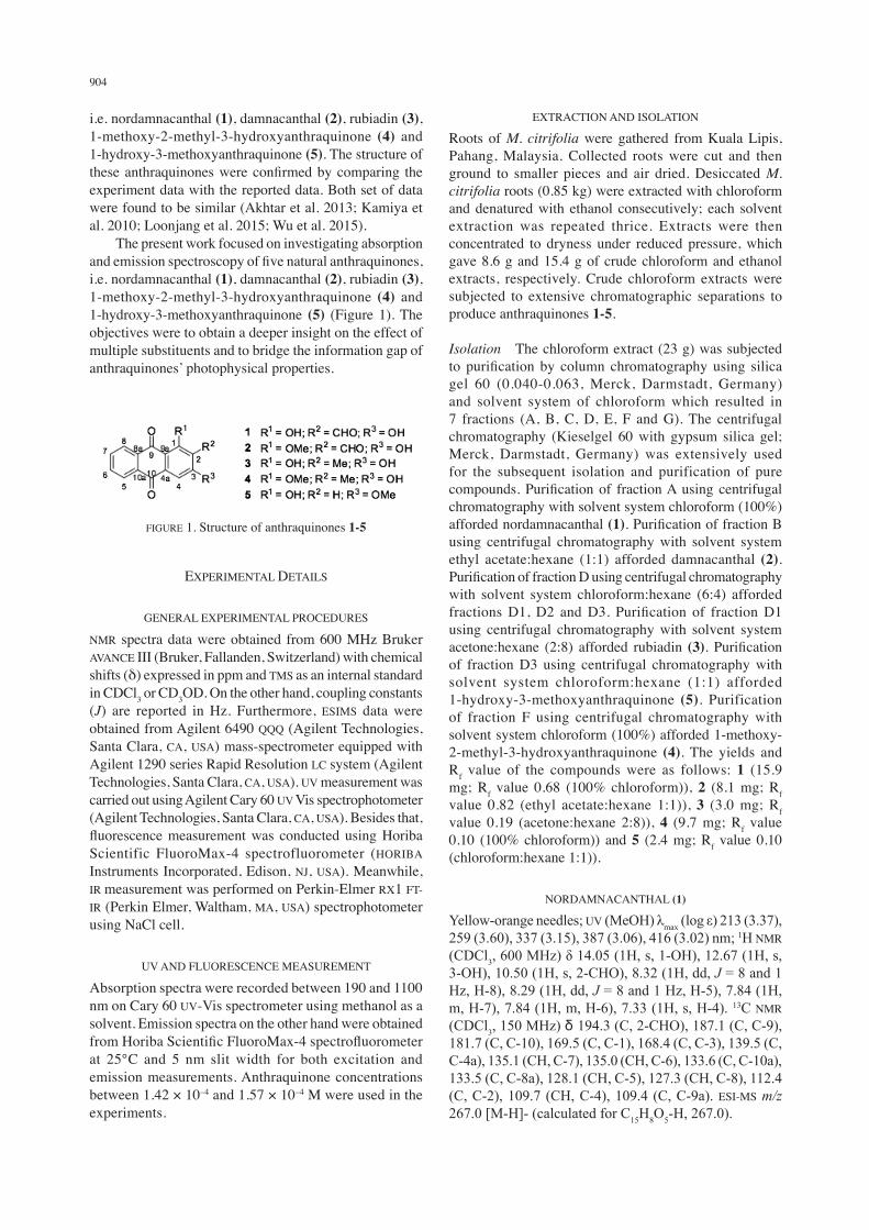
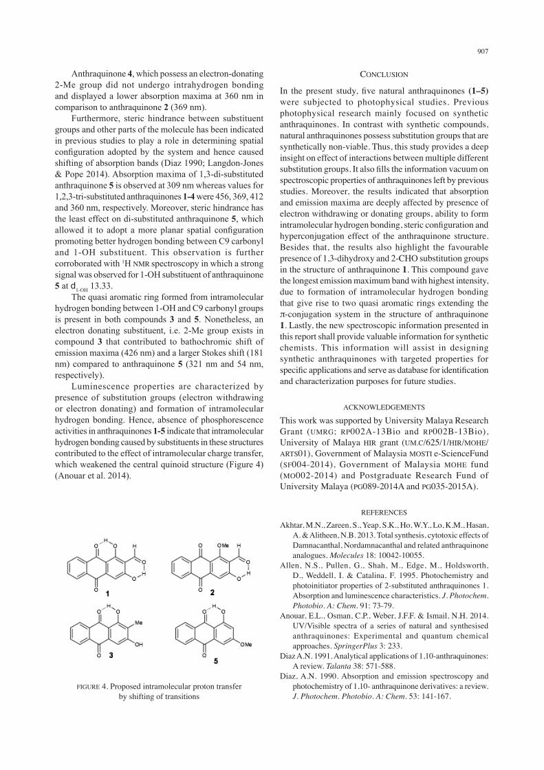
i.e. nordamnacanthal (1), damnacanthal (2), rubiadin (3), 1-methoxy-2-methyl-3-hydroxyanthraquinone (4) and 1-hydroxy-3-methoxyanthraquinone (5). The structure of these anthraquinones were confirmed by comparing the experiment data with the reported data. Both set of data were found to be similar (Akhtar et al. 2013; Kamiya et al. 2010; Loonjang et al. 2015; Wu et al. 2015). The present work focused on investigating absorption and emission spectroscopy of five natural anthraquinones, i.e. nordamnacanthal (1), damnacanthal (2), rubiadin (3), 1-methoxy-2-methyl-3-hydroxyanthraquinone (4) and 1-hydroxy-3-methoxyanthraquinone (5) (Figure 1). The objectives were to obtain a deeper insight on the effect of multiple substituents and to bridge the information gap of anthraquinones’ photophysical properties.
EXTRACTION AND ISOLATION
Roots of M. citrifolia were gathered from Kuala Lipis, Pahang, Malaysia. Collected roots were cut and then ground to smaller pieces and air dried. Desiccated M. citrifolia roots (0.85 kg) were extracted with chloroform and denatured with ethanol consecutively; each solvent extraction was repeated thrice. Extracts were then concentrated to dryness under reduced pressure, which gave 8.6 g and 15.4 g of crude chloroform and ethanol extracts, respectively. Crude chloroform extracts were subjected to extensive chromatographic separations to produce anthraquinones 1-5.
Isolation The chloroform extract (23 g) was subjected to purification by column chromatography using silica gel 60 (0.040-0.063, Merck, Darmstadt, Germany) and solvent system of chloroform which resulted in 7 fractions (A, B, C, D, E, F and G). The centrifugal chromatography (Kieselgel 60 with gypsum silica gel; Merck, Darmstadt, Germany) was extensively used for the subsequent isolation and purification of pure compounds. Purification of fraction A using centrifugal chromatography with solvent system chloroform (100%) afforded nordamnacanthal (1). Purification of fraction B using centrifugal chromatography with solvent system ethyl acetate:hexane (1:1) afforded damnacanthal (2). Purification of fraction D using centrifugal chromatography with solvent system chloroform:hexane (6:4) afforded fractions D1, D2 and D3. Purification of fraction D1 using centrifugal chromatography with solvent system acetone:hexane (2:8) afforded rubiadin (3). Purification of fraction D3 using centrifugal chromatography with solvent system chloroform:hexane (1:1) afforded 1-hydroxy-3-methoxyanthraquinone (5). Purification of fraction F using centrifugal chromatography with solvent system chloroform (100%) afforded 1-methoxy-2-methyl-3-hydroxyanthraquinone (4). The yields and Rf value of the compounds were as follows: 1 (15.9 mg; Rf value 0.68 (100% chloroform)), 2 (8.1 mg; Rf value 0.82 (ethyl acetate:hexane 1:1)), 3 (3.0 mg; Rf value 0.19 (acetone:hexane 2:8)), 4 (9.7 mg; Rf value 0.10 (100% chloroform)) and 5 (2.4 mg; Rf value 0.10 (chloroform:hexane 1:1)).
NORDAMNACANTHAL (1)
Yellow-orange needles; UV (MeOH) λmax (log ε) 213 (3.37), 259 (3.60), 337 (3.15), 387 (3.06), 416 (3.02) nm; 1H NMR (CDCl3, 600 MHz) δ 14.05 (1H, s, 1-OH), 12.67 (1H, s, 3-OH), 10.50 (1H, s, 2-CHO), 8.32 (1H, dd, J = 8 and 1 Hz, H-8), 8.29 (1H, dd, J = 8 and 1 Hz, H-5), 7.84 (1H, m, H-7), 7.84 (1H, m, H-6), 7.33 (1H, s, H-4). 13C NMR (CDCl3, 150 MHz) δ 194.3 (C, 2-CHO), 187.1 (C, C-9), 181.7 (C, C-10), 169.5 (C, C-1), 168.4 (C, C-3), 139.5 (C, C-4a), 135.1 (CH, C-7), 135.0 (CH, C-6), 133.6 (C, C-10a), 133.5 (C, C-8a), 128.1 (CH, C-5), 127.3 (CH, C-8), 112.4 (C, C-2), 109.7 (CH, C-4), 109.4 (C, C-9a). ESI-MS m/z 267.0 [M-H]- (calculated for C15H8O5-H, 267.0).
FIGURE 1. Structure of anthraquinones 1-5
EXPERIMENTAL DETAILS
GENERAL EXPERIMENTAL PROCEDURES
NMR spectra data were obtained from 600 MHz Bruker AVANCE III (Bruker, Fallanden, Switzerland) with chemical shifts (δ) expressed in ppm and TMS as an internal standard in CDCl3 or CD3OD. On the other hand, coupling constants (J) are reported in Hz. Furthermore, ESIMS data were obtained from Agilent 6490 QQQ (Agilent Technologies, Santa Clara, CA, USA) mass-spectrometer equipped with Agilent 1290 series Rapid Resolution LC system (Agilent Technologies, Santa Clara, CA, USA). UV measurement was carried out using Agilent Cary 60 UV Vis spectrophotometer (Agilent Technologies, Santa Clara, CA, USA). Besides that, fluorescence measurement was conducted using Horiba Scientific FluoroMax-4 spectrofluorometer (HORIBA Instruments Incorporated, Edison, NJ, USA). Meanwhile, IR measurement was performed on Perkin-Elmer RX1 FT-IR (Perkin Elmer, Waltham, MA, USA) spectrophotometer using NaCl cell.
UV AND FLUORESCENCE MEASUREMENT
Absorption spectra were recorded between 190 and 1100 nm on Cary 60 UV-Vis spectrometer using methanol as a solvent. Emission spectra on the other hand were obtained from Horiba Scientific FluoroMax-4 spectrofluorometer at 25°C and 5 nm slit width for both excitation and emission measurements. Anthraquinone concentrations between 1.42 × 10⁻⁴ and 1.57 × 10⁻⁴ M were used in the experiments.
905
DAMNACANTHAL (2)
Yellow amorphous; UV (MeOH) λmax (log ε) 203 (3.22), 248 (3.17), 279 (3.36), 316 (2.87), 379 (2.65) nm; 1H NMR (CDCl3, 600 MHz) δ 12.28 (1H, s, 3-OH), 10.47 (1H, s, 2-CHO), 8.30 (1H, dd, J = 8 and 1 Hz, H-8), 8.25 (1H, dd, J = 8 and 1 Hz, H-5), 7.83 (1H, td, J = 8 and 1 Hz, H-7), 7.78 (1H, td, J = 8 and 1 Hz, H-6), 7.68 (1H, s, H-4), 4.13 (1H, s, 1-OMe). 13C NMR (CDCl3, 150 MHz) δ 194.3 (C, 2-CHO), 182.3 (C, C-10), 180.5 (C, C-9), 167.0 (C, C-1), 166.9 (C, C-3), 142.0 (C, C-4a), 135.2 (C, C-8a), 135.2 (CH, C-7), 134.0 (CH, C-6), 132.8 (C, C-10a), 127.7 (CH, C-5), 127.4 (CH, C-8), 118.4 (C, C-2), 118.0 (C, C-9a), 113.4 (CH, C-4), 65.0 (C, 1-OMe). ESI-MS with m/z 281.0 [M-H]- (calculated for C16H10O5-H, 281.0).
RUBIADIN (3)
Yellow amorphous; UV (MeOH) λmax (log ε) 204 (3.63), 245 (3.31), 278 (3.42), 412 (2.78) nm; 1H NMR (CDCl3, 600 MHz) δ 13.11 (1H, s, 1-OH), 8.22 (1H, dd, J = 8 and 1 Hz, H-8), 8.15 (1H, dd, J = 8 and 1 Hz, H-5), 7.73 (1H, td, J = 8 and 1 Hz, H-7), 7.69 (1H, td, J = 8 and 1 Hz, H-6), 7.16 (1H, s, H-4), 2.14 (1H, s, 2-Me). 13C NMR (CDCl3, 150 MHz) δ 186.8 (C, C-9), 183.8 (C, C-10), 163.3 (C, C-1), 162.8 (C, C-3), 134.1 (CH, C-6), 134.5 (CH, C-7), 134.5 (C, C-8a), 133.6 (C, C-10a), 132.1 (C, C-4a), 127.2 (CH, C-5), 127.0 (CH, C-8), 119.2 (C, C-9a), 109.9 (C, C-2), 107.9 (CH, C-4), 8.2 (C, 2-Me). ESI-MS m/z 253.0 [M-H]- (calculated for C15H10O4-H, 253.1).
1-METHOXY-2-METHYL-3-HYDROXYANTHRAQUINONE (4)
Yellow needles; UV (MeOH) λmax (log ε) 205 (3.97), 239 (3.74), 278 (4.06), 360 (3.11) nm; 1H NMR (CDCl3, 600 MHz) δ 8.21 (1H, dd, J = 8 and 1 Hz, H-8), 8.12 (1H, dd, J = 8 and 1 Hz, H-5), 7.72 (1H, td, J = 8 and 1 Hz, H-7), 7.65 (1H, td, J = 8 and 1 Hz, H-6), 7.40 (1H, s, H-4), 3.84 (1H, s, 1-OMe), 2.21 (1H, s, 2-Me). 13C NMR (CDCl3, 150 MHz) δ 184.4 (C, C-10), 181.7 (C, C-9), 162.0 (C, C-3), 161.5 (C, C-1), 135.3 (C, C-8a), 134.6 (CH, C-7), 133.2 (CH, C-6), 132.8 (C, C-10a), 127.7 (C, C-2), 127.4 (CH, C-8), 126.6 (CH, C-5), 118.9 (C, C-9a), 109.7 (CH, C-4), 61.2 (C, 1-OMe), 9.2 (C, 2-Me). ESI-MS m/z 267.0 [M-H]- (calculated for C16H12O4-H, 267.1).
1-HYDROXY-3-METHOXYANTHRAQUINONE (5)
Yellow amorphous; UV (MeOH) λmax (log ε) 203 (3.65), 267 (2.57), 307 (2.05) nm; 1H NMR (CDCl3, 600 MHz) δ 13.33 (1H, s, 1-OH), 8.33 (1H, m, H-5), 8.33 (1H, m, H-8), 7.85 (1H, m, H-6), 7.85 (1H, m, H-7), 7.53 (1H, s, H-4), 6.26 (1H, s, H-2), 3.42 (1H, s, 3-OMe). ESI-MS m/z 253.0 [M-H] (calculated for C15H10O4-H, 253.1).
RESULTS AND DISCUSSION
In the present study, five natural anthraquinones, e.g. Nordamnacanthal (1), damnacanthal (2), rubiadin (3), 1-methoxy-2-methyl-3-hydroxyanthraquinone (4), and
1-hydroxy-3-methoxyanthraquinone (5) were isolated and subjected to absorption and emission spectroscopy studies. Past research on absorption and emission spectroscopy were mainly limited to anthraquinones of synthetic origins. Due to synthetic limitation, anthraquinone derivatives with substitution at positions -1, -4, -5, and -8 are more favoured as a result of activation by C9 carbonyl function of the anthraquinone structure (Langdon-Jones & Pope 2014). Nonetheless, unlike synthetic anthraquinones, the naturally occurring ones possess more diversity in substituent groups and positions. This is true even at the carbon position that is proven to be synthetically unfeasible, such as positions -2, -3, -6, and -7. Lastly, this is the first report on absorption and emission properties of natural anthraquinones 1-5.
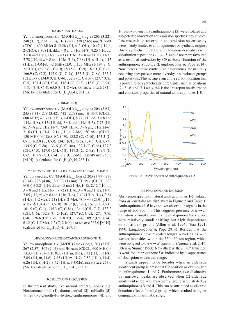
FIGURE 2. UV-Vis spectra of anthraquinones 1-5
ABSORPTION AND EMISSION
Absorption spectra of natural anthraquinones 1-5 isolated from M. citrifolia are displayed in Figure 2 and Table 1. Anthraquinones 1-5 have shown absorption signals in the range of 200-300 nm. This suggests presence of n → π* transition of fused aromatic rings and quinone backbones, with relatively small shifting but high dependence on substituent groups (Allen et al. 1995; Diaz 1991, 1990; Langdon-Jones & Pope 2014). Besides that, the anthraquinones have revealed longer wavelengths with weaker intensities within the 350-450 nm region, which were assigned to the n → π* transition (Anouar et al. 2014; Peters & Sumner 1953). Nevertheless, the n → π* transition is weak for anthraquinone 5 as indicated by disappearance of absorption within this range. Signals appear to be broader when an aldehyde substituent group is present at C2 position as exemplified in anthraquinones 1 and 2. Furthermore, two distinctive but narrower peaks are observed when C2-aldehyde substituent is replaced by a methyl group as illustrated by anthraquinones 3 and 4. This can be attributed as electron donation effect of methyl group, which resulted in higher conjugation in aromatic rings.
906
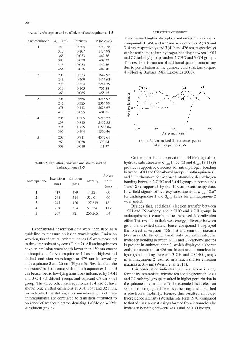
Experimental absorption data were then used as a guideline to measure emission wavelengths. Emission wavelengths of natural anthraquinones 1-5 were measured in the same solvent system (Table 2). All anthraquinones have an emission wavelength lower than 450 nm except anthraquinone 1. Anthraquinone 1 has the highest red shifted emission wavelength at 479 nm followed by anthraquinone 3 at 426 nm (Figure 3). Besides that, the emissions’ bathochromic shift of anthraquinones 1 and 3 can be ascribed to low-lying transitions influenced by 1-OH and 3-OH substituent groups and adjacent C9-carbonyl group. The three other anthraquinones 2, 4 and 5, have shown blue shifted emissions at 314, 354, and 321 nm, respectively. Blue shifting emission wavelengths of these anthraquinones are correlated to transition attributed to presence of weaker electron donating 1-OMe or 3-OMe substituent groups.
SUBSTITUENT EFFECT
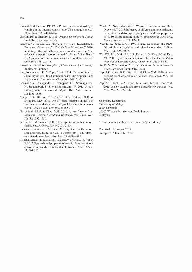
The observed higher absorption and emission maxima of compounds 1 (456 and 479 nm, respectively), 2 (369 and 314 nm, respectively) and 3 (412 and 426 nm, respectively) can be attributed to intrahydrogen bonding between 1-OH and C9 carbonyl groups and/or 2-CHO and 3-OH groups. This results in formation of additional quasi-aromatic ring due to perturbation in the quinone core structure (Figure 4) (Flom & Barbara 1985; Lakowicz 2006).
TABLE 1. Absorption and coefficient of anthraquinones 1-5
Anthraquinone λmax (nm) Intensity ε (M cm-1)1 241
313365387419456
0.2050.1070.0330.0300.0330.036
2749.261434.98442.56402.33442.56482.80
2 203248279316369
0.2330.2090.3240.1050.065
1642.921475.632284.39737.88455.15
3 204245278412
0.6680.3250.4130.095
4248.972064.992626.67601.05
4 205239278360
1.3850.8131.7250.194
9285.235452.8311566.841300.46
5 203267309
0.7110.0580.018
4517.61370.04111.37
TABLE 2. Excitation, emission and stokes shift of anthraquinones 1-5
AnthaquinoneExcitation
(nm)Emission
(nm)Intensity
Stokes shift (nm)
12345
419248245239267
479314426354321
17.12153.401127.61957.834256.265
606618111554
On the other hand, observation of 1H NMR signal for hydroxy substituents at d1-OH 14.05 (1) and d1-OH 13.11 (3) provides supportive evidence for intrahydrogen bonding between 1-OH and C9 carbonyl groups in anthraquinones 1 and 3. Furthermore, formation of intramolecular hydrogen bonding between 2-CHO and 3-OH groups in compounds 1 and 2 is supported by the 1H NMR spectroscopy data. Low field signals of hydroxy substituents at d3-OH 12.67 for anthraquinone 1 and d3-OH 12.28 for anthraquinone 2 were noted. Besides that, additional electron transfer between 1-OH and C9 carbonyl and 2-CHO and 3-OH groups in anthraquinone 1 contributed to increased delocalization effect. This resulted in the lowest energy difference between ground and exited states. Hence, compound 1 displayed the longest absorption (456 nm) and emission maxima (479 nm). On the other hand, only one intramolecular hydrogen bonding between 1-OH and C9 carbonyl groups is present in anthraquinone 3, which displayed a shorter emission maximum at 426 nm. In contrast, intramolecular hydrogen bonding between 3-OH and 2-CHO groups in anthraquinone 2 resulted in a much shorter emission maxima at 314 nm (Weislo et al. 2013). This observation indicates that quasi aromatic rings formed by intramolecular hydrogen bonding between 1-OH and C9 carbonyl groups resulted in higher perturbation in the quinone core structure. It also extended the p-electron system of conjugated heterocyclic ring and disturbed p-electron’s mobility. Hence, this resulted in lower fluorescence intensity (Weisstuch & Testa 1970) compared to that of quasi aromatic rings formed from intramolecular hydrogen bonding between 3-OH and 2-CHO groups.
FIGURE 3. Normalized fluorescence spectra of anthraquinones 1-5
907
Anthraquinone 4, which possess an electron-donating 2-Me group did not undergo intrahydrogen bonding and displayed a lower absorption maxima at 360 nm in comparison to anthraquinone 2 (369 nm). Furthermore, steric hindrance between substituent groups and other parts of the molecule has been indicated in previous studies to play a role in determining spatial configuration adopted by the system and hence caused shifting of absorption bands (Diaz 1990; Langdon-Jones & Pope 2014). Absorption maxima of 1,3-di-substituted anthraquinone 5 is observed at 309 nm whereas values for 1,2,3-tri-substituted anthraquinones 1-4 were 456, 369, 412 and 360 nm, respectively. Moreover, steric hindrance has the least effect on di-substituted anthraquinone 5, which allowed it to adopt a more planar spatial configuration promoting better hydrogen bonding between C9 carbonyl and 1-OH substituent. This observation is further corroborated with 1H NMR spectroscopy in which a strong signal was observed for 1-OH substituent of anthraquinone 5 at d1-OH 13.33. The quasi aromatic ring formed from intramolecular hydrogen bonding between 1-OH and C9 carbonyl groups is present in both compounds 3 and 5. Nonetheless, an electron donating substituent, i.e. 2-Me group exists in compound 3 that contributed to bathochromic shift of emission maxima (426 nm) and a larger Stokes shift (181 nm) compared to anthraquinone 5 (321 nm and 54 nm, respectively). Luminescence properties are characterized by presence of substitution groups (electron withdrawing or electron donating) and formation of intramolecular hydrogen bonding. Hence, absence of phosphorescence activities in anthraquinones 1-5 indicate that intramolecular hydrogen bonding caused by substituents in these structures contributed to the effect of intramolecular charge transfer, which weakened the central quinoid structure (Figure 4) (Anouar et al. 2014).
CONCLUSION
In the present study, five natural anthraquinones (1–5) were subjected to photophysical studies. Previous photophysical research mainly focused on synthetic anthraquinones. In contrast with synthetic compounds, natural anthraquinones possess substitution groups that are synthetically non-viable. Thus, this study provides a deep insight on effect of interactions between multiple different substitution groups. It also fills the information vacuum on spectroscopic properties of anthraquinones left by previous studies. Moreover, the results indicated that absorption and emission maxima are deeply affected by presence of electron withdrawing or donating groups, ability to form intramolecular hydrogen bonding, steric configuration and hyperconjugation effect of the anthraquinone structure. Besides that, the results also highlight the favourable presence of 1,3-dihydroxy and 2-CHO substitution groups in the structure of anthraquinone 1. This compound gave the longest emission maximum band with highest intensity, due to formation of intramolecular hydrogen bonding that give rise to two quasi aromatic rings extending the π-conjugation system in the structure of anthraquinone 1. Lastly, the new spectroscopic information presented in this report shall provide valuable information for synthetic chemists. This information will assist in designing synthetic anthraquinones with targeted properties for specific applications and serve as database for identification and characterization purposes for future studies.
ACKNOWLEDGEMENTS
This work was supported by University Malaya Research Grant (UMRG; RP002A-13Bio and RP002B-13Bio), University of Malaya HIR grant (UM.C/625/1/HIR/MOHE/ARTS01), Government of Malaysia MOSTI e-ScienceFund (SF004-2014), Government of Malaysia MOHE fund (MO002-2014) and Postgraduate Research Fund of University Malaya (PG089-2014A and PG035-2015A).
REFERENCES
Akhtar, M.N., Zareen, S., Yeap, S.K., Ho, W.Y., Lo, K.M., Hasan, A. & Alitheen, N.B. 2013. Total synthesis, cytotoxic effects of Damnacanthal, Nordamnacanthal and related anthraquinone analogues. Molecules 18: 10042-10055.
Allen, N.S., Pullen, G., Shah, M., Edge, M., Holdsworth, D., Weddell, I. & Catalina, F. 1995. Photochemistry and photoinitiator properties of 2-substituted anthraquinones 1. Absorption and luminescence characteristics. J. Photochem. Photobio. A: Chem. 91: 73-79.
Anouar, E.L., Osman, C.P., Weber, J.F.F. & Ismail, N.H. 2014. UV/Visible spectra of a series of natural and synthesised anthraquinones: Experimental and quantum chemical approaches. SpringerPlus 3: 233.
Diaz A.N. 1991. Analytical applications of 1,10-anthraquinones: A review. Talanta 38: 571-588.
Diaz, A.N. 1990. Absorption and emission spectroscopy and photochemistry of 1,10- anthraquinone derivatives: a review. J. Photochem. Photobio. A: Chem. 53: 141-167.
FIGURE 4. Proposed intramolecular proton transfer by shifting of transitions
908
Flom, S.R. & Barbara, P.F. 1985. Proton transfer and hydrogen bonding in the internal conversion of S1 anthraquinones. J. Phys. Chem. 89: 4489-4494.
Gordon, P.F. & Gregory, P. 1983. Organic Chemistry in Colour. Heidelberg: Springer Verlag.
Kamiya, K., Hamabe, W., Tokuyama, S., Hirano, K., Satake, T., Kumamoto-Yonezawa, Y., Yoshida, Y. & Mizushina, Y. 2010. Inhibitory effect of anthraquinones isolated from the Noni (Morinda citrifolia) root on animal A-, B- and Y-families of DNA polymerases and human cancer cell proliferation. Food Chemistry 188: 725-730.
Lakowicz, J.R. 2006. Principles of Fluorescence Spectroscopy. Baltimore: Springer.
Langdon-Jones, E.E. & Pope, S.J.A. 2014. The coordination chemistry of substituted anthraquinones: Developments and applications. Coordination Chem. Rev. 269: 32-53.
Loonjang, K., Duangjinda, D., Phongpaichit, S., Sawangjaroen, N., Rattanaburi, S. & Mahabusarakam, W. 2015. A new anthraquinone from Morinda elliptica Ridl. Nat. Prod. Res. 29: 1833-1838.
Madje, B.R., Shelke, K.F., Sapkal, S.B., Kakade, G.K. & Shingare, M.S. 2010. An efficient onepot synthesis of anthraquinone derivatives catalyzed by alum in aqueous media. Green Chem. Lett. Rev. 3: 269-273.
Nur Atiqah, M.N. & Choo, Y.M. 2016. A new flavone from Malaysia Borneo Marsdenia tinctoria. Nat. Prod. Res. 30(13): 1532-1536.
Peters, R.H. & Sumner, H.H. 1953. Spectra of anthraquinone derivatives. J. Chem. Soc. 0: 2101-2110.
Puenner, F., Schieven, J. & Hilt, G. 2013. Synthesis of fluorenone and anthraquinone derivatives from aryl- and aroyl-substituted propiolates. Org. Lett. 18: 4888-4891.
Seidel, N., Hahn, T., Liebing, S., Seichter, W., Kortus, J. & Weber, E. 2013. Synthesis and properties of new 9, 10-anthraquinone derived compounds for molecular electronics. New J. Chem. 37: 601-610.
Weisło, A., Niedziałkowski, P., Wnuk, E., Zarzeczan´ska, D. & Ossowski, T. 2013. Influence of different amino substituents in position 1 and 4 on spectroscopic and acid base properties of 9, 10-anthraquinone moiety. Spectrochim. Acta Mol. Biomol. Spectrosc. 108: 82-88.
Weisstuch, J. & Testa, A.C. 1970. Fluorescence study of 2-(N,N-Dimethylamino)pyridine and related molecules. J. Phys. Chem. 74: 2299-2302.
Wu, T.S., Lin, D.M., Shi, L.S., Damu, A.G., Kuo, P.C. & Kuo, Y.H. 2003. Cytotoxic anthraquinones from the stems of Rubia wallichiana DECNE. Chem. Pharm. Bull. 51: 948-950.
Xu, R., Ye, Y. & Zhao, W. 2010. Introduction to Natural Products Chemistry. Boca Raton: CRC Press.
Yap, A.C., Chan, K.G., Sim, K.S. & Choo, Y.M. 2016. A new oxolane from Enterobacter cloacae. Nat. Prod. Res. 30: 783-788.
Yap, A.C., Teoh, W.Y., Chan, K.G., Sim, K.S. & Choo Y.M. 2015. A new oxathiolane from Enterobacter cloacae. Nat. Prod. Res. 29: 722-726.
Chemistry DepartmentUniversity of MalayaJalan Universiti50603 Wilayah Persekutuan, Kuala LumpurMalaysia
*Corresponding author; email: [email protected]
Received: 21 August 2017Accepted: 5 December 2017