UNIVERSITI PUTRA MALAYSIA DETECTION OF HEPATITIS B … · HBcAg atau juga dikenali sebagai...
25
UNIVERSITI PUTRA MALAYSIA DETECTION OF HEPATITIS B CORE ANTIGEN USING A FUSION BACTERIOPHAGE SITI SALWA HASMONI. FBSB 2005 14
-
Upload
nguyencong -
Category
Documents
-
view
222 -
download
0
Transcript of UNIVERSITI PUTRA MALAYSIA DETECTION OF HEPATITIS B … · HBcAg atau juga dikenali sebagai...
UNIVERSITI PUTRA MALAYSIA
DETECTION OF HEPATITIS B CORE ANTIGEN USING A FUSION BACTERIOPHAGE
SITI SALWA HASMONI.
FBSB 2005 14
DETECTION OF HEPATITIS B CORE ANTIGEN USING A FUSION BACTERIOPHAGE
BY SIT1 SALWA HASMONI
Thesis Submitted to the School of Graduate Studies, Universiti Putra Malaysia, in Fulfilment of the Requirements of the Degree of Master of Science
September 2005
Specially dedicated to,
Papa and Mama, Kak Long, Kayna, Aji, Alol, Yan, and Limah
For their invaluable love, understanding, patience, support and care.
Abstract of thesis presented to the Senate Universiti Putra Malaysia in fulfilment of the requirement for the degree of Master of Science.
DETECTION OF HEPATITITS B CORE ANTIGEN USING A FUSION BACTERIOPHAGE
SIT1 SALWA BlNTl HASMONI
September 2005
Chairman: Associate Professor Tan Wen Siang, PhD
Faculty: Biotechnology and Biomolecular Sciences
Due to the many reported cases of hepatitis B disease around the world, a
keen interest among researches has aroused on the cause of the disease,
hepatitis B virus (HBV). One of the serological markers for HBV is hepatitis B
core antigen (HBcAg) that is a marker of the infectious material and it is the
most accurate index of the viral replication. The importance of the HBcAg
especially when considering the close relationship with the viral DNA load
has created revolutionary studies on the HBcAg ever since. The HBV
nucleocapsid or HBcAg is extremely immunogenic during infection and after
immunization. A fusion bacteriophage that interacts with HBcAg has been
isolated from a phage display peptide library. The phage interacts tightly to
HBcAg and thus has the potential to be further developed as a diagnostic
reagent. In this study, two immunoassays have been developed using the
fusion bacteriophage to detect HBcAg. Phage-ELISA and phage-dot blot
assay could detect not only purified HBcAg but also HBcAg in serum
samples. As low as 10 ng of HBcAg can be significantly detected by 1 012
pfulml of fusion phage when the reading at 405 nm was measured (hO5 =
0.4). Using the fusion bacteriophage, these newly developed immunoassays

provide an easier and cheaper option for detecting HBcAg. The sensitivity of
these immunoassays demonstrates the potential and perhaps vast future
uses to detect HBcAg. The fusion phage is also capable of purifying the
HBcAg due to its capability to precipitate HBcAg.
Abstrak
PEAWsT* SULTAN ADUL SAMAD ~ W T I WTRA MAJAY%
tesis yang dikemukakan kepada Senat Universiti Putra Malaysia sebagai memenuhi keperluan untuk ijazah Master Sains
PENGESANAN ANTIGEN TERAS VIRUS HEPATITIS B DENGAN MENGGUNAKAN BAKTERIOFAJ REKOMBINAN
Oleh
SIT1 SALWA BlNTl HASMONI
September 2005
Pengerusi: Profesor Madya Tan Wen Siang, PhD
Bioteknologi dan Sains Biomolekul
Terdapat banyak kes penyakit Hepatitis B telah dilaporkan. Ini menimbulkan
minat yang tinggi kepada para penyelidik untuk menjalankan penyelidikan
tentang punca penyakit tersebut iaitu virus Hepatitis B (HBV). Salah satu
penunjuk serologi virus ini adalah HBcAg. la merupakan penanda jangkitan
HBV yang sangat efektif dan paling tepat dalam menunjukkan kehadiran
aktiviti replikasi virus tersebut. Kepentingan HBcAg terutama mengenai
hubungan rapat protein tersebut dengan jumlah DNA HBV telah mencetus
revolusi di dalam bidang penyelidikan yang telah memberi kesan yang
mendalam. HBcAg atau juga dikenali sebagai nukleokapsid HBV adalah
sangat imunogenik semasa jangkitan dan selepas immunisasi. Satu
bakteriofaj rekombinan yang telah dipilih daripada sebuah perpustakaan
pameran faj didapati dapat berinteraksi kuat dengan HBcAg. Faj yang
membawa peptide-peptida tertentu ini berpotensi untuk dijadikan suatu
reagen dalam bidang diagnostik. Di akhir pengajian ini, dua jenis imunoasai
yang menggunakan bakteriofaj rekombinan untuk mengesan HBcAg telah
berjaya dihasilkan. Faj-ELISA dan asai faj-dot blot mampu mengesan HBcAg
yang telah ditulenkan dan juga HBcAg di dalam sample-sampel serum.
Sejumlah lo1* pfulml faj rekombinan berupaya mengesan dengan baik 10 ng
HBcAg apabila bacaan tindakbalas yang diambil pada gelombang 405 nm
diukur (ko5 = 0.4). Imunoasai-imunoasai baru ini menyediakan suatu kaedah
yang lebih murah dan mudah dalam mengesan HBcAg disamping
mempamerkan pengesanan HBcAg yang sensitif. Oleh yang demikian,
imunoasai-imunoasai ini berpotensi tinggi untuk digunakan di masa hadapan
dalam mengesan HBcAg khususnya. Faj rekombinan ini juga boleh
digunakan untuk menulenkan HBcAg, berdasarkan kebolehannya
memendakkan HBcAg.
ACKNOWLEDGEMENTS
I would like to express my deepest appreciation and thanks to my main
supervisor, Assoc. Prof. Dr. Tan Wen Siang, for his never ending guidance
and encouragement throughout this project. His patience and dedication has
inspired me to give my very best in completion of this study and the thesis.
My sincere thanks to my co-supervisor, Prof. Datin Dr. Khatijah Yusoff for her
invaluable advice and constructive criticism during the entire progress of this
project. Her suggestions and comments definitely helped in the success of
this thesis.
A special note of appreciation also goes out to the staff from Faculty of
Biotechnology and Biomolecular Studies and everyone who have contributed
in one way or another to this study.
I am truly indebted to my lab mates, Kak Tan, Thong Chuan, Kah Fai,
Andrew, Swee Tin, Taznim, Yan Peng, Lalita, Kak Fieda, Kak Onie, Abg. Zul,
Max, Watti, Kak Sue, Kak Raha and last but not least Dr. Majid Eshaghi for
their helpful collaboration and discussion. Also not forgetting my friends,
Deela, Nurul, Elia, Fazu, Surini, Che Lina, Mok, Shahrul, Tajul, Lan, and a
bunch of others that have really helped me in any possible way there could
be. Last but not least, my heartiest appreciation to Soffiyan and his family for
their continual support and understanding for me to proceed completion.
May god bless you all.
vii
I certify that an Examination Committee met on 3oth September 2005 to conduct the final examination of Siti Salwa Hasmoni on her Master of Science thesis entitled "Detection of Hepatitis B Core Antigen Using a Fusion Bacteriophagen in accordance with Universiti Pertanian Malaysia (Higher Degree) Act 1980 and Universiti Pertanian Malaysia (Higher Degree) Regulations 1981. The Committee recommends that the candidate be awarded the relevant degree. Members of the Examination Committee are as follows:
Janna Ong Abdullah, PhD Lecturer Faculty of Biotechnology and Biomolecular Sciences Universiti Putra Malaysia (Chairman)
Raja Noor Zaliha Raja Abdul Rahman, PhD Associate Professor Faculty of Biotechnology and Biomolecular Sciences Universiti Putra Malaysia (Internal Examiner)
Muhajir Hamid, PhD Lecturer Faculty of Biotechnology and Biomolecular Sciences Universiti Putra Malaysia (Internal Examiner)
Sheila Nathan, PhD Associate Professor Faculty of Science and Technology Universiti Teknologi Malaysia (External Examiner)
Date: 25 OCT ZO@j
. . . Vlll
This thesis submitted to the Senate of Universiti Putra Malaysia and has been accepted as fulfilment of the requirement for the degree of Master of Science. The members of the Supervisory Committee are as follows:
Tan Wen Siang, PhD Associate Professor Faculty of Biotechnology and Biomolecular Sciences Universiti Putra Malaysia (Chairman)
Khatijah Yusoff, PhD Professor Faculty of Biotechnology and Biomolecular Sciences Universiti Putra Malaysia (Member)
AlNl IDERIS, PhD ProfessorIDean School of Graduate Studies Universiti Putra Malaysia
DECLARATION
I hereby declare that the thesis is based on my original work except for quotations and citations which have been duly acknowledged. I also declare that it has not been previously or concurrently submitted for any other degree at UPM or other institutions.
SIT1 SALWA HASMONI
TABLE OF CONTENTS
DEDICATION ABSTRACT ABSTRAK ACKNOWLEDGEMENTS APPROVAL DECLARATION LlST OF TABLES LlST OF FIGURES LlST OF ABBREVIATION
CHAPTER
INTRODUCTION Introduction
LITERATURE REVIEW
Page ii iii v vii viii X
xv xvi xix
Hepatitis B Virus (HBV) 2.1 . I Hepatitis B Virus Classification Biology of Hepatitis B Virus 2.2.1 HBV Morphology 2.2.2 Genomic Organisation 2.2.3 Replication of Hepatitis B Virus 2.2.4 Viral Proteins Immunology of Hepatitis B Virus in Human 2.3.1 Serological markers Detection of Hepatitis B Virus 2.4.1 Production of Monoclonal Antibodies 2.4.2 Radioimmunoassays 2.4.3 lmmunoassays 2.4.4 Molecular Techniques
2.4.4.1 PCR Biology of Filamentous Bacteriophage 2.5.1 Morphology of Filamentous
Bacteriophage 2.5.2 Genomic Organisation and the
Products of Filamentous Phage 2.5.3 Replication of Filamentous
Bacteriophage 21 Phage Display Technology 22 2.6.1 Phage Display System 23 Application of Phage Display Technology 24 Principles of Affinity Chromatography 26 2.8.1 Coupling Gels for Ligand lmmobilisation 27
MATERIALS AND METHODS Materials 3.1 .I Standard Solutions and Buffers 3.1 . I Liquid and Solid Media 3.1.2 Serum Samples Preparation and Purification of Full-Length and Truncated HBcAg 3.2.1 The Bradford Assay 3.2.2 SDS-PAGE 3.2.3 Western Blot Analysis Preparation and Purification of Fusion M I 3 Bacteriophage 3.3.1 Phage Titration 3.3.2 Large Scale preparation and
Purification of Fusion M I 3 Bacteriophage 36 3.3.2.1 Extraction of ssDNA 38 3.3.2.2 Agarose Gel Electrophoresis 38 3.3.2.3 DNA Sequencing 39
Detection of HBV Serum Samples with PCR 40 3.4.1 Extraction of HBV DNA 40 3.4.2 PCR 4 0 Pretreatment of Serum Samples 4 1 Phage-Enzyme-Linked lmmunoabsorbent Assay (Phage-ELISA) 4 1 3.6.1 Optimization of the Concentrations of
HBcAg, Fusion Phage, Temperatures, and Blocking Buffers 42
Enzyme-Linked lmmunoabsorbent Assay (ELISA) Phage-ELISA for the Detection of HBcAg in Serum Samples Phage-Dot Blot Assay 3.9.1 Optimization of the Concentrations of
Fusion Phage, HBcAg, and Anti-HBcAg Monoclonal Antibodies
Dot Blot Assay Phage-Dot Blot Assay for the Detection of HBcAg in Serum Samples Specificity Test of Phage-ELISA and Phage-Dot Blot Assay for the Detection of HBcAg using Fusion Phage 3.1 2.1 Phage-ELISA 3.1 2.2 Phage-Dot Blot Assay Phage-Precipitation Assay 3.1 3.1 lmmobilisation
(Cross-Linking Optimization) 3.1 3.2 Precipitation of Full-Length and
Truncated HBcAg 3.1 4 Purification of Full-Length and Truncated
xii
HBcAg using Fusion M I 3 Bacteriophage
RESULTS Preparation, Purification and Analysis of Full-length and Truncated HBcAg Large Scale Preparation and Purification of Fusion M I 3 Bacteriophage 4.2.1 Cesium Chloride Ultracentrifugation Analysis of HBV Samples with PCR Phage-Enzyme-Linked lmmunoabsorbent Assay (Phage-ELISA) 4.4.1 Optimization of the Concentrations of
HBcAg, Fusion Phage, Temperatures, And Blocking Buffers
Enzyme-Linked lmmunoabsorbent Assay (EL I SA) Phage-ELISA for the detection of HBcAg in Serum Samples Phage-Dot Blot Assay 4.7.1 Optimization of the Concentrations of
the Fusion Phage, HBcAg, and Anti-HBcAg Monoclonal Anti bodies
Dot Blot Assay Phage-Dot Blot Assay for the Detection of HBcAg in Serum Samples Specificity Test of Phage-ELISA and Phage-Dot Blot Assay for the Detection of HBcAg using Fusion Phage Phage-Precipitation Assay 4.1 1 .I Optimization of the Cross-Linking
Reaction 4.1 1.2 Precipitation of Full-Length and
Truncated HBcAg
DISCUSSION Preparation, Purification and Analysis of Full-length and Truncated HBcAg Large Scale Preparation and Purification of Fusion M I 3 Bacteriophage Analysis of HBV Samples with PCR Phage-Enzyme-Linked lmmunoabsorbent Assay (Phage-ELISA) Enzyme-Linked lmmunoabsorbent Assay (ELISA) Phage-ELISA for the detection of HBcAg in Serum Samples Phage-Dot Blot Assay Dot Blot Assay Phage-Dot Blot Assay for the Detection of HBcAg in Serum Samples
. . . X l l l
5.1 0 Specificity Test of Phage-ELISA and Phage-Dot Blot Assay for the Detection of HBcAg using Fusion Phage
5.1 1 Phage-Precipitation Assay 5.1 2 Purification of Full-Length and Truncated
HBcAg using fusion MI3 bacteriophage
SUMMARY AND CONCLUSION
REFERENCES BlODATA OF THE AUTHOR PUBLICATIONS
xiv
LIST OF TABLES
Table
3.1 Standard solutions and buffers
3.2 Parameters and conditions for the first part of Phage-
Page
30
43
ELlSA
Parameters and conditions towards the final optimization 44
of Phage-ELISA
Parameters and conditions for the optimization of Phage- 47
dot blot assay
The number of blue plaque formed or the number of
uncoupled ligands after the cross-linking reaction
Final Optimized Parameters and Conditions of Phage- 116
ELI SA
Final Optimized Parameters and Conditions for Phage-Dot 116
Blot Assay
LIST OF FIGURES
Figure
2.1
2.2
A schematic representative of virion structure of HBV
Genome organisation of HBV
Interaction of different cell system in the immune
response against HBV
A schematic representative of Ff bacteriophage
The genome of the M I 3
Principle of affinity chromatography
SDS-PAGE of full-length and truncated HBcAg
fractionated on sucrose gradient centrifugation
SDS-PAGE and Western blot of concentrated full-length 57
and truncated HBcAg
Plaque assay showing blue recombinant M I 3 plaques
Agarose gel electrophoresis of ssDNA fusion M I 3
bacteriophage
Chromatogram of the nucleotide sequence of the fusion 61
M I 3 phage clone WSFFSNI
Detection of HBV serum samples using the PCR 62
Detection of HBcAg using the fusion phage 64
Optimization of detection of HBcAg using different set of 66
concentration of fusion phage
Detection of different range of amount of HBcAg using
the fusion phage
Page
6
7
xvi
Detection of a narrower range of HBcAg using the fusion 68
phage
Graph representing the detection of HBcAg using 1.0 x 70
10" to 1.0 x 1 012 pfulml of the fusion phage with milk
diluent as the blocking buffer
4.12(a) Graph presenting the detection of HBcAg using 1.0 x
10" to 1.0 x 1 012 pfulml of the fusion phage that uses
SEA blocking solution as the blocking buffer
4.1 2(b) Detection of HBcAg using 1.0 x 10" to 1.0 x 10¹² pfulml 71
of fusion phage with different blocking duration
Optimization of the coating duration for the detection of 73
HBcAg using fusion phage (1.0 x 10'' to 1.0 x 10¹²
pfulml)
Optimised detection of HBcAg using fusion phage via
Phage-ELISA
Detection of HBcAg with anti-HBcAg monoclonal
antibody via ELlSA
Detection of HBcAg in HBV positive serum samples
using fusion phage via phage-ELISA
Optimization of the blocking buffer for the detection of
HBcAg using fusion phage
Optimization of different duration and temperature of
HBcAg incubation for phage-dot blot assay
Optimization of the duration and temperature of anti-
xvii
HBcAg incubation for phage-dot blot assay
Optimization of the dilutions of anti-HBcAg mAb (primary 82
antibody) for phage-dot blot assay
Detection of HBcAg with fusion phage via dot blot assay 84
Detection of HBcAg with anti-HBcAg monoclonal 85
antibody via dot blot assay
Detection of HBcAg in HBV positive serum samples with 86
fusion phage via dot blot assay
Specificity test results for phage-ELISA 88
The specificity test phage-dot blot results that correspond 89
to that of the phage-ELISA
Specificity test results for phage-ELISA
Specificity test phage-dot blot assay results
SDS-PAGE of precipitated products using the fusion
phage immobilized on CNBr activated-agarose as ligand
Western blotting of precipitated products with anti-HBcAg 96
antibody
xviii
CHAPS
CNBr
BSA
CCC
CTL
c-terminus
DC
DNA
LIST OF ABBREVIATIONS
encapsidation signal
alpha
beta
microgram (1 0-6 g)
microlitre (1 0-6 I)
micromolar M)
ampicillin
antibody to HBcAg
antibody to HBeAg
antibody to HBsAg
antibody to HBxAg
degree centigrade
3-[(3-chol-amidopropy1)-dimethylammonio]-l -
propanesulfonate
cyanogen bromide
base pair
bovine serum albumin
cytosinelcore
covalently closed circular
cytotoxic T lymphocyte
carboxyl terminus
dendritic cell
deoxy-ribonucleic acid
xix
DNase
dNTP
DTT
d s
EDTA
HBcAg
HBeAg
HBsAg
HBV
HBxAg
HCC
HCI
I FN
IFN-y
I L2
I L4
I L5
lL l0
IL12
H IV
HLA
IgG
Deoxyribonuclease
deoxynucleoside triphosphate
1,4-dithiothreitol
Double stranded
ethyler/ediaminetetraacetic acid
enzyme-linked immunoabsorbent assay
Gram
Hour
hepatitis B core antigen
hepatitis B e antigen
hepatitis B surface antigen
hepatitis B virus
hepatitis B x antigen
hepatocellular carcinoma
hydrochloric acid
Interferon
interferon gamma
interleukin 2
interleukin 4
interleukin 5
interleukin 10
interleukin 12
human immunodeficiency virus
human leukocyte antigen
immunoglobulin G
IgM
l PTG
kb
kDa
K ~ ' ~ '
I
LB
L-HBsAg
M
m g
MgC12
M-HBsAg
min
mm
mRNA
Na Cl
NDV
NK
nM
immunoglobulin M
isopropyl-P-d-thiogalactopyranoside
kilobase
kilodalton
relative dissociation constant
Litre
Luria Bertani
large surface antigen
Molar
monoclonal antibody
milligram (1 om3 g)
magnesium chloride
medium surface antigen
Minute
millilitre (1 0-3 I)
milimeter (1 0-3 m)
messenger ribonucleic acid
sodium chloride
Newcastle disease virus
natural killer
nanomolar (1 0-' M)
nanometer ( I 0-' m)
nucleotide(s)
amino terminus
optical density
xxi
ORF
P
PAGE
PBS
PCR
PEG
pf u
P ~ R N A
preC
preC/C
preS
preS/S
PreS1
PreS2
RF
RNA
RNase
SDS
S-HBsAg
ssDNA
TAE
open reading frame
polymerase protein
polyacrylamide gel electrophoresis
phosphate buffered saline
Polymerase Chain Reaction
polyethylene glycol
plaque forming unit
pregenomic RNA
precore
precore and core
hepatitis B preS genes
preS and surface
N-terminal region of L-HBsAg comprising 108 or 11 9
amino acid
region of M and L-HBsAg comprising 55 amino acid
replicative form
ribonucleic acid
ribonuclease
revolutions per minute
room temperature
second
sodium dodecyl sulphate
small surface antigen
single stranded DNA
tris acetate EDTA buffer
xxii
Taq
TBE
TBS
TE
TEMED
Thl
Th2
TP
USA
v
v/v
WHO
w/v
x g
X-gal
thermus aquaticus thermostable DNA
tris-buffered EDTA solution
tris-buffered saline
tris-EDTA buffer
tetramethyl ethylenediamine
T helper 1
T helper 2
terminal protein
United States of America
Volt
volume/volume
World Health Organization
weightlvolume
centrifugal force
5-bromo-4-chloro-3-indol-P-D-galactosidase
xxiii
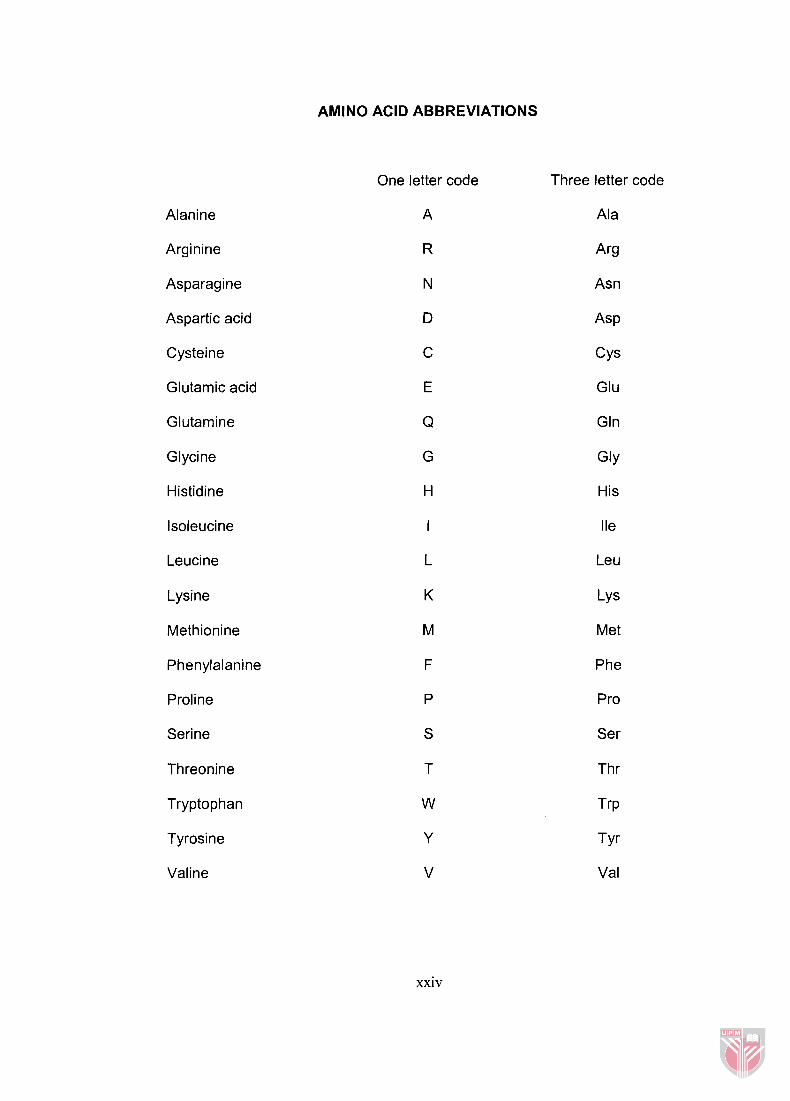
Alanine
Arginine
Asparagine
Aspartic acid
Cysteine
Glutamic acid
Glutamine
Glycine
Histidine
lsoleucine
Leucine
Lysine
Methionine
Phenylalanine
Proline
Serine
Threonine
Tryptophan
Tyrosine
Valine
AMINO ACID ABBREVIATIONS
One letter code Three letter code
Ala
Arg
Asn
Asp
CYS
Glu
Gln
G ~ Y
His
I le
Leu
LY s
Met
Phe
Pro
Ser
Thr
T ~ P
T Y ~
Val
xxiv


![[Presentasi]Line Detection Using Hough Transform](https://static.fdokumen.site/doc/165x107/5571fe7049795991699b6284/presentasiline-detection-using-hough-transform.jpg)

![Lp Hepatitis II[1]](https://static.fdokumen.site/doc/165x107/563dbb54550346aa9aac3a49/lp-hepatitis-ii1.jpg)