UNIVERSITI PUTRA MALAYSIA SEROLOGICAL SURVEY OF … · Kuda-kuda terlibat tidak pernah di dalam...
25
UNIVERSITI PUTRA MALAYSIA SEROLOGICAL SURVEY OF EQUINE INFECTIOUS ANEMIA VIRUS (EIAV) IN HORSES IN SELANGQR ABDUL NASSER ASHOUR ALTAEB FPV 2004 21
Transcript of UNIVERSITI PUTRA MALAYSIA SEROLOGICAL SURVEY OF … · Kuda-kuda terlibat tidak pernah di dalam...

UNIVERSITI PUTRA MALAYSIA
SEROLOGICAL SURVEY OF EQUINE INFECTIOUS ANEMIA VIRUS (EIAV) IN HORSES IN SELANGQR
ABDUL NASSER ASHOUR ALTAEB
FPV 2004 21

SEROLOGICAL SURVEY OF EQUINE INFECTIOUS ANEMIA VIRUS (EIAV) IN HORSES IN SELANGQR
ABDUL NASSER ASHOUR ALTAEB
A project paper submitted to Faculty of Veterinary Medicine, Universiti Putra Malaysia in Partial Fulfillment of the Requirements for the Degree of Master of
Veterinary Medicine (M.V.M)
UNIVERSITI PUTRA MALAYSIA 43400 SERDANG, SELANGOR DARUL EHSAN
MALAYSIA
March 2004
DEDICATION
To MY PARENTS, MY WIFE, MY SON HAMAM AND MY BROTHERS
AND SISTERS

ABSTRACT
An abstract of the project paper presented to the Faculty of Veterinary Medicine, Universiti Putra Malaysia in partial fulfillments of the requirements
for the degree of Master of Veterinary Medicine
SEROLOGICAL SURVEY OF EQUINE INFECTIOUS ANEMIA VIRUS (EIAV) IN HORSES IN SELANGOR
BY
ABDUL NASSER ASHOUR ALTAEB
March 2004
Supervisor: Dr. Mohd Azam Khan Goriman Khan
Faculty: Veterinary Medicine
A study was carried out to determine the prevalence of Equine Infectious Anemia
Virus (EIAV) among horses of various clubs in the state of Selangor. A total of 94
serum samples was obtained from horses of various breeds, sex and age and tested
for the presence of antibodies against EIAV using; (i) competitive enzyme-linked
immunosorbent assay (CELISA) and (ii) agar gel immunodiffusion (AGID) test, to
demonstrate the presence of antibodies against p26 recombinant gag protein. All
horses sampled in this study were kept in stables and have access to paddock grazing.
Horses have no history of contact with imported stocks. In this study none of the
sera had antibodies against EIA virus. It is possible that the strict import policies
imposed, and the almost non-existent importation of horses for the purpose of
breeding has prevented the introduction of the disease into Malaysia.

ABSTRAK
Abstrak daripada kertas projek yang dibentangkan kepada Fakulti Perubatan Veterinar, Universiti Putra Malaysia adalah sebahagian
daripada keperluan memenuhi Ijazah Sarjana dalam Perubatan Veterinar
KAJI SELIDIK SEROLOGI TERHADAP VIRUS ANEMIA BERJANGKIT KUDA (EIAV) KE ATAS KUDA-KUDA DI SELANGOR
Oleh
ABDUL NASSER ASHOUR ALTAEB
Mac 2004
Penyelia : Dr. Mohd Azam Khan Goriman Khan
Fakulti: Perubatan Veterinar
Satu kajian telah dijalankan untuk mengkaji prevalens virus anaemia berjangkit
ekuin (EIAV) di kalangan kuda-kuda dari berbagai kelab di negeri Selangor.
Sejumlah 94 sampel serum telah diambil dari kuda-kuda pelbagai baka, jantina dan
umur, dan telah diuji untuk menentukan kehadiran antibodi-antibodi terhadap EIAV
dengan menggunakan (i) asai imunoerap terangkai enzim bersaing (CELISA) dan
(ii) ujian imunoresapan gel agar-agar (AGID) untuk menunjukkan kehadiran
antibodi-antibodi terhadap rekombinan protein gag p26. Semua kuda-kuda yang di
sampel dalam kajian ini disimpan di dalam kandang dan dibenarkan melakukan
ragutan alongan. Kuda-kuda terlibat tidak pernah di dalam sejarah bersentuh dengan
stok-stok yang di import. Di dalam kajian ini tiada satu pun serum-serum yang diuji
mengandungi antibodi terhadap virus EIA. Kemungkinan polisi-polisi ketat
pengimportan yang dikuatkuasakan dan hampir tiadanya pengimportan kuda-kuda
untuk tujuan pembiakan, telah mencegah kemasukan penyakit tersebut ke Malaysia.

APPROVAL
It is hereby certified that we have read this project paper entitled "Serological Survey
of Equine Infectious Anemia Virus (EIAV) in Horses in Selangor" by Abdul Nasser
Ashour Altaeb and in our opinion; it is satisfactory in terms of scope, quality and
presentations as fulfillment of the requirement for the degree of Master of Veterinary
Medicine, VPD 5908 project.
Dr. MOHD AZAM KHAN GORIMAN KHAN DVM (UPM), PhD (Glasgow)
Lecturer Faculty of Veterinary Medicine
Universiti Putra Malaysia (Supervisor)
ASSOC. PROF. Dr. ABDUL RAHMAN OMAR DVM (UPM), PhD (Cornell)
Lecturer Faculty of Veterinary Medicine
Universiti Putra Malaysia (Co-Supervisor)

ACKNOWLEDGEMENTS
I thank Allah for the big support he has provided me during this research effort. I
would like to express my sincere gratitude and appreciation to my supervisor, Dr.
Mohd Azam Khan Goriman Khan and my co-supervisor Associate Professor Dr.
Abdul Rahman Omar, for their valuable concern, guidance, advice, and support
through this research.
I would also like to express my appreciation to Associate Professor Dr. Bashir
Ahmed for his recommendation and providing the CELISA Kit used in this study. I
would also like to express my gratitude to Prof. Dato Dr. Sheikh Omar A. Rahman
for his advices, recommendations, and always support.
I would also like to thank Dr. Noraniza, and Mr. Salehuddin for helping to collect my
samples.
My sincere thanks are also extended to the entire faculty; post graduate students, my
friends, and technicians at Biologics Laboratory.
Finally, my appreciation and my utmost gratitude are expressed to my wife, for her
support and encouragement and to my parents for their prayers.

DECLARATION
I hereby declare that the project paper is based on my original work except
quotations and citations, which have been duly acknowledged. I also declare that it
has not been previously or concurrently submitted for any degree at UPM or other
institutions.
Abdul Nasser Ashour Altaeb Date.. .
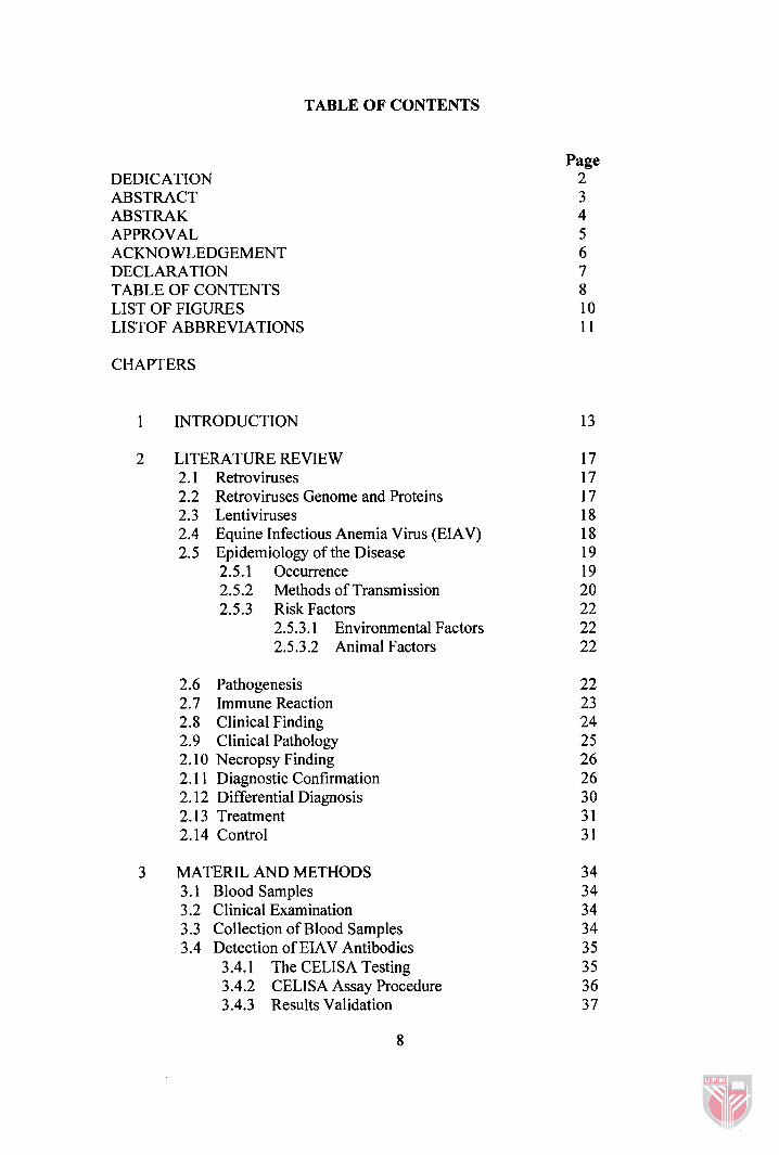
TABLE OF CONTENTS
DEDICATION ABSTRACT ABSTRAK APPROVAL ACKNOWLEDGEMENT DECLARATION TABLE OF CONTENTS LIST OF FIGURES LISTOF ABBREVIATIONS
CHAPTERS
1 INTRODUCTION
2 LITERATURE REVIEW 2.1 Retroviruses 2.2 Retroviruses Genome and Proteins 2.3 Lentiviruses 2.4 Equine Infectious Anemia Virus (EIAV) 2.5 Epidemiology of the Disease
2.5.1 Occurrence 2.5.2 Methods of Transmission 2.5.3 Risk Factors
2.5.3.1 Environmental Factors 2.5.3.2 Animal Factors
2.6 Pathogenesis 2.7 Immune Reaction 2.8 Clinical Finding 2.9 Clinical Pathology 2.10 Necropsy Finding 2.1 1 Diagnostic Confirmation 2.12 Differential Diagnosis 2.13 Treatment 2.14 Control
MATERIL AND METHODS 3.1 Blood Samples 3.2 Clinical Examination 3.3 Collection of Blood Samples 3.4 Detection of EIAV Antibodies
3.4.1 The CELISA Testing 3.4.2 CELISA Assay Procedure 3.4.3 Results Validation
Page 2 3 4 5 6 7 8 10 11

3.4.4 Interpretation of Result 3.4.5 The AGID Test 3.4.6 Preparation of Agar Gel 3.4.7 Cutting Wells in the Agar 3.4.8 AGID Assay Procedure 3.4.9 Interpretation of Results
4 RESULTS 4.1 CELISA 4.2 AGID
5 DISCUSSION
6 CONCLUSION AND SUGGESTIONS
REFERENCES APPENDIX 1 APPENDIX 2 VITAE

LIST OF FIGURES
FIGURES
CELISA PLATE LAYOUT
CELISA KIT REAGENTS
CUTTING WELLS IN THE AGAR GEL
AGID TEST RESULT
PAGE

LIST OF ABBREVIATIONS
AIDS
BIV
CA
CAEV
CDC
CF12h
CF
ED
EIAV
FEA
FEK
FIV
gP
HIV
HI
ID
IN
LTR
MA
NC
OIE
Acquired immune deficiency syndrome
Bovine immunodeficiency virus
Mature capsid
Caprine arthritis encephalitis virus or Visna virus
Center for disease control
Canine osteosarcoma cells
Complement fixation test
Equine dermal cell line
Equine infectious anemia virus
Feline embryonic cells
Fetal equine kidney cell
Feline immunodeficiency virus
Fluorescence polarization test
Glycoprotein
Human immunodeficiency virus
Hemagglutination inhibition
Immunnodiffussion test
Integrase
Long terminal repeats
Matrix
Nucleocapsid
Office International des Epizooties

PCR
R T
SIVS
SN
su
TM
u3
u5
WHO
WY
Polymerase chain reaction
Reverse transcriptase
Simian immunodeficiency viruses
Serum neutralization test
Env gene encodes the surface
Transmembrane
Unique 3
Unique 5
World Health Organization
Wyoming strain

CHAPTER 1
INTRODUCTION
Equine infectious anemia (EIA) or Swamp Fever is a multisystemic retroviral
disease of equidae, characterized by hemolytic anemia that is immune mediated
(Clabough, 1990). The disease is caused by Equine infectious anemia virus (EIAV),
a member of the lentivirus subfamily of retroviruses. The virus possesses the surface
glycoproteins, gp90 and gp45 and four major nonglycosylated internal core proteins,
known as p26, p15, p l l and p9 (Parekh et al., 1980; Montelaro et al., 1984; Hussain
et al., 1988) (Appendix 2).
The disease is distributed worldwide in the Equidae family; and has been
reported in horses, donkeys and mules (Montelaro et al., 1984; Hammond et al.,
1997). Once an animal is infected, it will remain infected for the rest of its life
(Montelaro et al., 1984; Clements and Zink, 1996; Issel et al., 1982).
After infection, the animal experiences three distinct disease phases: acute,
chronic and inapparent carrier (Clabough, 1990; Roberts and Lucas, 1987). The acute
stage of infection is characterized by recurrent febrile episodes with high virus load.
After resolution of the primary viremia, most animals develop chronic EIA, which is
characterized by recurrent cycles of the disease, associated with weight loss and
anemia, leading to a prolonged and symptomatic period, and finally an inapparent
carrier stage for the rest of the animal's life (Hammond et al., 1997).

Most infected horses develop a vigorous humoral immune response, the
envelope glycoproteins being the major immunogens. Each episode of viremia is
associated with the appearance of a new and predominant antigenic variant of the
virus; caused by point mutations in the env gene that codes the viral surface proteins
(Montelaro et al., 1984).
Although EIA infection persists life-long, during non-fibrile intervals the
likelihood of isolating EIA from blood samples is close to nil. The diagnosis of EIA
rests entirely on serological evidence (Issel et al., 1988). Coggins et al., (1972)
developed an agar gel immunodifision (AGID) test to detect precipitating
antibodies to the core protein p26, which is known to be group-reactive and
antigenically stable (Payne et al., 1984). The AGID test is the only test officially
recognized by the Office International des Epizooties (01E) in Paris (Pearson and
Coggins, 1979). A positive AGID test noted 15 to 25 days post infection (Coggins et
al., 1972), confirms that the virus is present as infected horses produce antibodies to
EIAV proteins within 12 days of infection (Issel and Cook, 1993).
Enzyme-linked immunosorbent assay (ELISA) was developed in the late
1980's for faster and more sensitive serodiagnosis (Issel et al., 1988; Archambault et
al., 1989; Ramachandran et al., 1990). Competitive ELISA (CELISA) was
developed using purified antigens from cell culture adapted EIAV strain Wyoming,
to detect antibodies against p26 protein. Although the authors found a good
correlation between AGID and CELISA, the latter could not detect weak positive
antibody titers (Burki et al., 1992; Issel and Cook, 1993). Another ELISA was
developed using the amino-terminal portion of a recombinant gp45 protein as antigen

(Hancock and Tsang, 1986; Archambault et al., 1989; Thomas et al., 1991; Lew et
al., 1993). Soutullo et al., (2001) designed an ELISA test using linear and cyclic
synthetic peptides from gp45 and gp90 as antigen. To date, the largest amount of
serological information relating to the presence of EIAV infection has been obtained
using the internal core protein p26 and surface glycoprotein gp90 and gp45 isolates
as the antigens.
Since clinical diagnosis is difficult for acute and inapparent infection, the
movement of horses across borders stands the risk of causing economic
embarrassments and interference to sporting events due to EIA. Economic losses
due to the disease can be large as there is no treatment or vaccination, for this
disease. Mortality rates may approach 50% and the athletic ability of horses
reduced.
Serologic evidence for EIAV infection has been reported in many countries
around the world. Using AGID and CELISA the prevalence of this disease was 1.5-
2.5% in the USA, 6% in Canada, a low level in France, 1.6% in West Germany, and
15-25% in Argentina and 50% in Brazil. The disease has never been recorded in
Malaysia. Large-scale movements of horses during wartime have been responsible
for extensive dissemination of the disease. At the present time there is another surge
of infection in most countries, possibly due to obligatory testing conducted as
surveillance of the disease. It is suggested that the rapid expansion of 'pleasure
horses' activity in affluent countries leads to more movement and opportunities for
spread of the infection from relatively few donors (Coggins, 1984).
Since there are no records on EIAV seroprevalence among horses in
Malaysia, the objective of this study was to investigate the seroprevalence of Equine
Infectious Anemia (EIA) among horses in Selangor using two different methods,
Competitive Enzyme Linked Imunosorbent Assay and Agar Gel Immunodiffusion
(Coggins Test).
CHAPTER 2
LITERATURE REVIEW
2.1 Retroviruses
In 1981 the center for disease control (CDC) USA, first described the
symptoms of disease that would be identified later as acquired immune deficiency
syndrome (AIDS). AIDS caused by a retrovirus infect other animals such as rodents,
monkeys, and horses. Nineteen years after the isolation of the human
immunodeficiency virus (HIV), the etiology agent of AIDS, the World Health
Organization (WHO) estimates 65 million people to be infected with HIV world
wide, thus establishing HIV as the leading cause of death by infectious agents.
The impact of HIV has greatly changed the direction of scientific research
and brought the field of retrovirology to the forefront of medicine. The Retroviridae
family includes viruses with a diploid RNA genome that utilizes reverse
transcriptase, an RNA dependent DNA polymerase. Retroviruses are enveloped
viruses ranging in size from 80-100 nm in diameter and contain the enzymes reverse
transcriptase integrase and protease (Coffin et al., 1997).
2.2 Retrovirus Genome and Proteins
As reviewed by Coffin et al., (1997) the retrovirus family can be divided in two
main groups, based on genome organizations, simple and complex; Simple
retroviruses have gag, pro, pol and env genes that encode polyproteins. The gag
gene encodes the mature capsid (CA), matrix (MA), and nucleocapsid (NC)
17

structural proteins. The pol encodes reverse transcriptase (RT), and integrase (IN).
The gag and pol polyproteins are proteolyticlally cleaved via viral protease, encoded
by the Pro gene. The env gene encodes the surface (SU) and transmembrane (TM)
outer structure proteins. The env polyprotein is cleaved by host cell proteases. The
retrovirus provirus also contains non-coding long terminal repeats (LTR) located at
the 5'and 3' ends. The LTR consists of U3 (unique 3') and U5 regions. The U3
element occurs once at 3' end of viral genomic RNA and twice in proviral DNA.
The U3 region is between 120 - I200 nucleotides in length.
2.3 Lentiviruses
The term lentivirus originates from the Latin world lenti meaning slow. This
sub group of retroviruses is named due to the long incubation period from initial
infection with the virus, to appearance of disease symptoms. Viruses in the
lentiviruses group of retroviruses are the human immunodeficiency virus (HIV-1 and
HIV-2), simian immunodeficiency viruses (SIVS), equine infectious anemia virus
(EIAV), bovine immunodeficiency virus (BIV), visna virus or caprine arthritis
encephalitis virus (CAEV), and feline immunodeficiency virus (FIV). EIAV and
other lentiviruses encods a dUTPase enzyme (designated DU), which maps to the pol
gene (McClure et al., 1988: Elder et al., 1992).
2.4 Equine Infectious Anemia Virus (EIAV)
In vitro, several strains of EIAV have been shown to infect and replicate in
feline embryonic cells (FEA), canine osteosarcoma cells (CF12h), and equine
macrophages. Strains of EIAV exhibit differing cellular tropism. The Wyoming
strain (WY) is a highly virulent wild type strain of EIAV, and its replication in vitro
is restricted to equine macrophages in which it replicates to high titers. The WY
strain was adapted to replicate in ED, FEK, FEA and CF12h cell lines and was
termed the Malmquist strain (Malmquist et al., 1973). EIAV differs from other
human and non-human lantiviruses in vivo in that it has not been found to infect
lymphocytes; it is predominantly macrophage tropic (Clabough, 1990; Issel et al.,
1988). Pathogenically, EIA differs in several respects from other lentivirus
infections, in that it does not induce direct immunosuppression (Montelaro et aI.,
1989), but circulating immune-complexes (Henson et al., 1973).
2.5 Epidemiology of the Disease
2.5.1 Occurrence
EIA has been diagnosed in several different continents. In Europe, it is most
prevalent in the northern and central regions. It has appeared in most states in the
United States and the provinces of Canada but the principal enzootic areas are the
Gulf Coast region and the northern wooded sections of Canada (Paquette, 1985).
Diagnosis of the disease was made in Australia in 1959, but the incidence appears to
be very low (Lepherd, 1981). The only area of Australia in which EIA could be
regarded as being endemic is along the inland river systems of central and western
Queensland. In a serological survey in this area in 1978, 21.7% of horses and 23%
of properties were positive for EIAV (Thomas and Elder, 1978). The disease was
also reported once in Thailand in 1996 and Mongolia (Vallat, 2003) (Appendix 1).
The morbidity varies considerably and depends on the strain of the virus, and
the inoculum delivered by the biting insects (Coggins, 1984). Extensive serological
surveys over large areas, using the agar gel immunodiffusion (AGID) Coggins test
have shown the prevalence rates, ranging from 1.5 - 2.5 % in the United States, to
19
50% in Brazil (Jenner and Romulo, 1994). The prevalence of infection varies
depending on the population of horses, the proportion of carrier and the density of
insect vectors (Hall et al., 1988). Large-scale movements of horses during wartime
have been responsible for extensive dissemination of the disease. The possible
detection of the infection at present is due to obligatory testing carried out. Rapid
expansion of 'pleasure horse' activity in affluent countries may lead to more
movement of horses and opportunities for spread of the infection from relatively few
donors (Coggins, 1984).
2.5.2 Methods of Transmission
EIA virus is relatively resistant to most disinfectants and cannot be destroyed
by boiling for 15 minutes, but is destroyed by sunlight. It persists for several months
at room temperature in urine, feces, dried blood and serum. It is present in all
tissues, secretions and excretions and may persist in the body for up to 18 years,
providing a source of infection for most of the animal's life (Issel et al., 1988).
Horses that are febrile and show clinical signs of EIA have a higher titer of viremia
and much more likely to serve as a source of disease transmission than inapparent
carriers (Kono, 1969; Issel et al., 1982; Orrego, 1983).
Infection by the EIA virus usually occurs by transmission of blood between
infected and uninfected horses, although body secretions may contain virus and
could serve as a source of virus (Tshjian et al., 1984). Short - term contact is
usually insufficient to cause spread, but continued, close association
with susceptible animals usually results in infection. Spread within a group is slow,
although occasionally fairly rapid spread is observed in large groups of horses
assembled at racetracks or army depots (Clabough, 1990).
20

Insect vectors, the primary mode of transmission, with disease passage
occurring through interrupted feeding of stable fly (Stomoxys calcitrans), deer flies
(Chrysops spp.) and horse flies (Tabanus spp. and Hypomirta spp.) (Stein et al.,
1942; Hawkins et al., 1973, 1976; Kemen et al., 1978; Cupp and Kemen, 1980; Issel
et al., 1982; Foil et al., 1983; Issel and Foil, 1984). The disease spreads most actively
in summer and in marshy or wooded areas, suggesting that bites by insects may be
the most important method of spread. It is probable that transmission is mechanical
only (Cupp and Kemen, 1980; Issel and Foil, 1984). Therefore, wild type EIAV will
not multiply in any species of insects (Shen et al., 1978; Williams et al., 1981).
The virus has been transmitted under experimental and natural conditions from
inapparently infected animals (Issel et al., 1982; Foil et al., 1988) where viremia
titters are substantially lower than those observed in horses with clinical EIA (Kono
et a1 1976; Issel et al., 1982). In enzootic areas, outbreaks have been caused by the
use of untreated biological preparations of equine origin (Issel et al., 1988).
Kemen and Coggins (1972) have reported intrauterine transmission. Foals
have reportedly become infected through the milk of infected dams, but relatively
large amounts of virus must be ingested to cause infection and the digestive tract is
not a major portal of entry. Foals of infected dams are less susceptible to natural
infection than adults possibly due partly to persistence of colostral antibodies (Foil et
al., 1983). The opposite can also happen and the passively immune foal develops a
fatal case of the acute disease (Issel et al., 1985).

2.5.3 Risk Factors
2.5.3.1 Environmental Factors
There are marked seasonal incidence of the disease, most cases occurring in
the summer and autumn. This is associated with low-lying and newly settled bush
area and due to greater number of the insect vectors in such areas (Issel et al., 1992).
Distances between horses are important to the carrier. Although tabanids are strong
flyers, they usually prefer to complete an interrupted meal on the initial host or a
nearby host (Foil, 1983). Prolonged therapy with corticosteroids and poor
environment or management is known to induce recrudescence of EIA (Kono et al.,
1976).
2.5.3.2 Animal Factors
All breeds and age groups of equidae are susceptible to EIAV. The
indigenous Criollo horses of Argentina are reported to be much more resistant to
infection and only mildly affected by the disease (Shen et al., 1984). Foals have
lower tabanid burdens than older horses due in part to their more vigorous defense
movements (Issel et al., 1985; Foil et al., 1985).
2.6 Pathogenesis
After infection EIAV multiplies in tissues that have abundant macrophages,
notably the liver, spleen, lymph nodes, lungs and kidney (Issel et al., 1988; Clabough
1990) as viral replication occurs only in mature tissue of macrophages and does not
occur in circulating monocytes (Maury 1994; Sellon et al., 1992). The acute form of
the disease is thought to be associated with massive virus replication in and
destruction of macrophages, but the actual cause of death is unknown. There is good

evidence that the vascular lesions and the erythrocyte fragility are part of an immune
reaction (Clabough 1990; Issel et al., 1988).
2.7 Immune Reaction
The immune response to EIAV is responsible for controlling replication of
the virus and also plays an important role in the pathogenesis of the disease. The
major clinical signs and lesions of EIA are attributable to the host response to the
virus and not direct viral damage to the tissue (Sellon, 1993). Replication of EIAV
stimulates a strong immune response that is detectable within 7 to 10 days of
infection. Antibodies to the p26 core protein are detectable by AGID test in almost
all horses 45 days after infection and by 60 days after infection antibodies to gp45
and gp90 are present (Clabough, 1990). The immune response includes the
production of virus neutralizing antibodies, complement-fixating antibodies, and
cytotoxic T lymphocytes (McGuire et al., 1994). The immune responses are
responsible for the termination of viremia, but this effect is not mediated by
antibody-dependent cellular cytotoxicity against EIAV- infected macrophages
(Tschetter et al., 1997). Virus-antibody complex are readily phagocytosed by cells
of the reticulo-endothelial system, including tissue macrophages, and are involved in
the development of the fever, depression, thrombocytopenia, anemia and
glumerulonephritis, which are characteristic of the disease (Sellon, 1993).
Infection with EIAV shortens the lifespan of circulating red blood cells to
about 38 days (McGuire et al., 1969). EIAV also have suppressive effect on
erythroid series cell in bone marrow (Swardson et al., 1992). The anemia caused by
EIAV is not wholly due to the immune response of the host, but with severe

combined immunodeficiency of the infection (Perryman et al., 1988). EIAV does not
affect megakaryocytes and the suppressive effect of infection is due to at least in part
to alpha and beta tumor necrosis factors (Crawford et al., 1996).
2.8 Clinical Findings
An incubation period of two to four weeks is usual in natural outbreaks of
equine infectious anemia. Outbreaks usually follow a pattern of slow spread to
susceptible horses after the introduction of an infected animal. After infection, the
equine experiences three distinct disease phases: acute, chronic and inapparent
carrier (Roberts and Lucas, 1987).
Occasionally the initial attack is mild and may be followed by rapid clinical
recovery. As a rule there is initial anorexia, depression, profound weakness, and loss
of condition. Ataxia is a prominent sign in many cases and in some is recorded as the
only clinical abnormality (McClure et al., 1982). There is intermittent fever (up to
41 "C), which may rise and fall rapidly, sometimes varying as much as 1 "C within 1
hour. Jaundice, oedema of the ventral abdomen, the prepuce and legs, and petechial
hemorrhages in the mucosa, especially under the tongue and in the conjunctiva, may
be observed. Pallor of the mucosa does not occur in this early stage and they tend to
be congested and oedematous (Clabough, 1990).
There is a characteristic increase in rate and intensity of the heart sounds,
which are greatly exacerbated by moderate exercise. Myocarditis, manifested by
tachycardia and arrhythmia, are described as being diagnostic. Respiratory signs are
not marked, there is no dyspnea until the terminal stages, but there may be a thin
24