Crusty Leaf Spot Disease of Mango
3
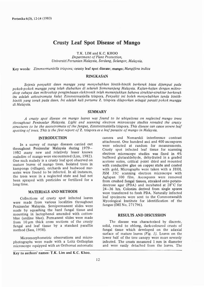
Pertanika 6(3), 12-14 (1983) Crusty Leaf Spot Disease of Mango T.K. LIM and K.C. KHOO Department of Plant Protection, Universiti Pertanian Malaysia, Serdang, Selangor, Malaysia. Key words: Zimmermaniella trispora; crusty leaf spot disease; mango; Mangifera indica RINGKASAN Sejenis penyakit daun mangga yang menyebabkan bintik-bintik berkerak biasa dijumpai pada pokok-pokok mangga yang telah diabaikan di seluruh Semenanjung Malaysia. Kajian-kajian dengan mikro- skop cahaya dan mikroskop pengimbasan elektronik telah menunjukkan bahawa struktur-struktur berkerak itu adalah askostromata kulat Zimmennaniella trispora. Penyakit ini boleh menyebabkan tanda bintik- bintik yang teruk pada daun. Ini adalah kali pertama Z. trispora dilaporkan sebagai parasit pokok mangga di Malaysia. SUMMARY A crusty spot disease on mango leaves was found to be ubiquitous on neglected mango trees throughout Peninsular Malaysia. Light and scanning electron microscope studies revealed the crusty structures to be the ascostromata of the fungus, Zimmennaniella trispora. This disease can cause severe leaf spotting of trees. This is the first report of z. trispora as a leaf parasite of mango in Malaysia. INTRODUCTION In a survey of mango diseases carried out throughout Peninsular Malaysia during 1979- 19B2 many new and relatively lesser known maladies of mango were encountered (Lim, 1982). One such malady is a crusty leaf spot observed on mature leaves of mango trees. Isolated trees in kampongs (villages), orchards and budwood nur- series were found to be infected. In all instances, the trees were in a neglected state and had not been sprayed with pesticides or fertilized for a long time. MATERIALS AND METHODS Collections of crusty spot infected leaves were made from various localities throughout Peninsular Malaysia. Semipermanent slides were made by squashing the hard fungal tissue and mounting in lactophenol amended with cotton- blue (aniline blue). Penn anent slides were made from 10 Mm thick cross sections of the crusty fungal and leaf tissue by a standard paraffin method (Sass, 1958). Micromorphometric observations and micro- photographs were made with a Leitz Orthoplan microscope equipped with an Orthomat automatic Key to authors' names: T.K. Lim and K.C. Khoo. 12 camera and Nomarskii interference contrast attachment. One hundred asci and 400 ascospores were selected at random for measurements. Crusty spot infected leaf tissue for scanning electron microscope studies was fixed in 4% buffered glutaraldehyde, dehydrated in a graded acetone series, critical point dried and mounted with conductive glue on copper stubs and coated with gold. Micrographs were taken with a JEOL JSM 35C scanning electron microscope with Agfapan 100 film. Ascospores were removed from crushed fungal tissues, streaked onto potato- dextrose agar (PDA) and incubated at 28° C for 24-36 hrs. Colonies derived from single spores were transferred to fresh PDA. Naturally infected leaf specimens were sent to the Commonwealth Mycological Institute for identification of the fungus (lMI No.2 71796). RESULTS AND DISCUSSION The disease was characterised by discrete, solid, round to oblong, dark-coloured crusts of fungal tissue which developed on the adaxial surface of mature leaves (Fig. 1). Leaves on the lower half of the tree canopy were more severely infected. The crusts measured 1 mm in diameter and were easily detached from the leaves. The
Transcript of Crusty Leaf Spot Disease of Mango
Pertanika 6(3), 12-14 (1983)
Crusty Leaf Spot Disease of Mango
T.K. LIM and K.C. KHOODepartment ofPlant Protection,
Universiti Pertanian Malaysia, Serdang, Selangor, Malaysia.
Key words: Zimmermaniella trispora; crusty leaf spot disease; mango; Mangifera indica
RINGKASAN
Sejenis penyakit daun mangga yang menyebabkan bintik-bintik berkerak biasa dijumpai padapokok-pokok mangga yang telah diabaikan di seluruh Semenanjung Malaysia. Kajian-kajian dengan mikroskop cahaya dan mikroskop pengimbasan elektronik telah menunjukkan bahawa struktur-struktur berkerakitu adalah askostromata kulat Zimmennaniella trispora. Penyakit ini boleh menyebabkan tanda bintikbintik yang teruk pada daun. Ini adalah kali pertama Z. trispora dilaporkan sebagai parasit pokok manggadi Malaysia.
SUMMARY
A crusty spot disease on mango leaves was found to be ubiquitous on neglected mango treesthroughout Peninsular Malaysia. Light and scanning electron microscope studies revealed the crustystructures to be the ascostromata of the fungus, Zimmennaniella trispora. This disease can cause severe leafspotting of trees. This is the first report of z. trispora as a leaf parasite of mango in Malaysia.
INTRODUCTION
In a survey of mango diseases carried outthroughout Peninsular Malaysia during 197919B2 many new and relatively lesser knownmaladies of mango were encountered (Lim, 1982).One such malady is a crusty leaf spot observed onmature leaves of mango trees. Isolated trees inkampongs (villages), orchards and budwood nurseries were found to be infected. In all instances,the trees were in a neglected state and had notbeen sprayed with pesticides or fertilized for along time.
MATERIALS AND METHODS
Collections of crusty spot infected leaveswere made from various localities throughoutPeninsular Malaysia. Semipermanent slides weremade by squashing the hard fungal tissue andmounting in lactophenol amended with cottonblue (aniline blue). Pennanent slides were madefrom 10 Mm thick cross sections of the crustyfungal and leaf tissue by a standard paraffinmethod (Sass, 1958).
Micromorphometric observations and microphotographs were made with a Leitz Orthoplanmicroscope equipped with an Orthomat automatic
Key to authors' names: T.K. Lim and K.C. Khoo.
12
camera and Nomarskii interference contrastattachment. One hundred asci and 400 ascosporeswere selected at random for measurements.Crusty spot infected leaf tissue for scanningelectron microscope studies was fixed in 4%buffered glutaraldehyde, dehydrated in a gradedacetone series, critical point dried and mountedwith conductive glue on copper stubs and coatedwith gold. Micrographs were taken with a JEOLJSM 35C scanning electron microscope withAgfapan 100 film. Ascospores were removedfrom crushed fungal tissues, streaked onto potatodextrose agar (PDA) and incubated at 28° C for24-36 hrs. Colonies derived from single sporeswere transferred to fresh PDA. Naturally infectedleaf specimens were sent to the CommonwealthMycological Institute for identification of thefungus (lMI No.2 71796).
RESULTS AND DISCUSSION
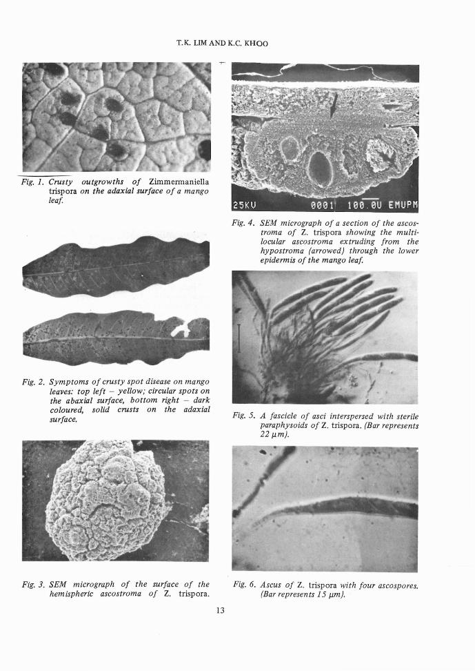
The disease was characterised by discrete,solid, round to oblong, dark-coloured crusts offungal tissue which developed on the adaxialsurface of mature leaves (Fig. 1). Leaves on thelower half of the tree canopy were more severelyinfected. The crusts measured 1 mm in diameterand were easily detached from the leaves. The
T.K. LIM AND K.C. KHOO
Fig. 1. Crusty outgrowths of Zimmennaniellatrispora on the adaxial surface of a mangoleaf,
Fig. 2. Symptoms of crusty spot disease on mangoleaves: top left - yellow; circular spots onthe abaxial surface, bottom right - darkcoloured, solid crusts on the adaxialsurface.
Fig. 3. SEM micrograph of the surface of thehemispheric ascostroma of Z. trispora.
13
Fig. 4. SEM micrograph of a section of the ascostroma of Z. trispora showing the multilocular ascostroma extruding from thehypostroma (arrowed) through the lowerepidermis of the mango leaf,
Fig. 5. A fascicle of asci interspersed with sterileparaphysoids of Z. trispora. (Bar represents22 11m).
Fig. 6. Ascus of Z. trispora with four ascospores.(Bar represents 15 11m).
CRUSTY LEAF SPOT DISEASE OF MANGO
fungus also gave rise to small, yellow, circularspots about 2-3 mm in diameter, each with adark, necrotic centre on the opposite, abaxialsurface of infected leaves (Fig. 2). Often, thiscrusty spot disease occurred on leaves which wereinfected with sooty mould and epiphytic greenalgae.
The structures were the erumpent ascostromata of the ascomycetous fungus, Zimmermaniellatrispora P. Henn. (Dothideaceae Dothideales).The crusty structure had a rough, convolutedsurface with some discontinuous deep fissures(Fig. 3). The fungus stroma originated in themesophyll and proliferated through the lowerepidermis forming a sessile, hemispheric, erumpentportion continuous with the hypostroma (Fig. 4).The ascostromata were multilocular and thelocules were obovate to oval and bore fasciclesof clavate, hyaline asci interspersed with sterile,filiform paraphysoids (Fig. 5). The mean dimensions of an ascus. from the point of attachmentto the apex were 104.37 Jim (range 72.00 145.00 Jim) and 4.80 Jim (range 4.76-4.92 Jim)at its widest width. The ascus bore three, sometimes four hyaline, ellipsoid ascospores (Fig. 6).Ascus with three ascospores and ascus with fourascospores occurred in the frequency ratio of97:3. The ascospore measured 18.94 Jim (range15.00-24.00 Jim) and 4.70 Jim (range 4.684.74 Jim) on the average. Non-sporulating greyishwhite colonies were obtained from single ascespores on PDA.
This report represents the first record ofZ. trispora as a leaf parasite of mango. There isa dearth of information on Zimmermaniella as aleaf parasite; however, much more is known aboutBagnisiopsis, a similar and closely related ascomycetous leaf parasite on Melastomaceae (Petrak,1928; Miller and Barton, 1943: Shirakawa, 1956).
14
The Widespread occurrence of Zimmermaniellacrusty spot on mango throughout PeninsularMalaysia suggests that the disease is not a recentone. That the disease has not been previouslydescribed is probably because little emphasis hasbeen given to local fruit disease research in thepast.
The survey indicated that the disease couldresult in severe leaf spotting of whole trees.Nevertheless, the disease could be considered tobe of minor importance especially when compared to other leaf disease like anthracnose causedby Colletotrichum gloeosporioides.
ACKNOWLEDGEMENTS
Grateful acknowledgements are due to Dr.D. W. Minter of the Commonwealth MycologicalInstitute for identifying the fungus and to thetechnical staff of the Electron Microscopy Unit,University of Agriculture Malaysia.
REFERENCES
LIM, T.K. (1982): Maladies of mango in Malaysia. XXIInternational Horticulture Congress, 29th Aug-4thSept. 1983, Hamburg, Fed. Republic of Germany.(Abstr. 1367).
MILLER, 1.H. and BARTON, M.G. c1943): Study ofBagnisiopsis species on the Melastomaceae. Mycologia, 35: 312-334.
PETRAK, F. VON, (1920): Uber Bagnisiopsis und verwandte Gattungen. Hedwigia, 68: 251-290.
SASS, 1.E., (1958): Botanical microtechnique. Ames.Iowa State Univ. Press.
SHIRAKAWA, H.S. (1956): The development of Cocostroma (Bagnisiopsis) nuda. A mer. 1. Bot., 43:372-377.
(Received 15 August 1983)