ULTRASTRUCTURAL LOCALIZATION OF M1 FUSION · PDF fileJurnal Biosains, 17(2), 9–20, 2006...
12
Jurnal Biosains, 17(2), 9–20, 2006 ULTRASTRUCTURAL LOCALIZATION OF M1 FUSION PROTEIN ON EQUATORIAL SEGMENT OF ACROSOME INTACT AND FREE/REACTED HAMSTER SPERMATOZOA 1 Mahanem Mat Noor* and 2 Moore H D M 1 School of Biosciences and Biotechnology, Faculty of Science and Technology, Universiti Kebangsaan Malaysia, 43600 Bangi, Selangor, Malaysia 2 Department of Molecular Biology and Biotechnology, University of Sheffield, S10, 2UH, United Kingdom Abstrak: Kajian telah dijalankan untuk menentukan kedudukan antigen M1 serta faktor yang mempengaruhi pengekspresan terhadap sperma hamster yang telah mengalami tindak balas akrosom secara fisiologi dan bukan fisiologi dengan mengaitkan keputusan pemerhatian melalui mikroskop cahaya dan elektron. Kaedah kajian melibatkan penyediaan sperma hamster dengan akrosom dan tanpa akrosom atau telah mengalami tindak balas akrosom. Dalam penyediaan sampel sperma, setiap sampel perlakuan menjalani kaedah pewarnaan imunopendarfluor dan pelabelan imunoemas secara selari. Sperma tanpa akrosom atau telah mengalami tindak balas akrosom masing-masing disediakan melalui prosedur mekanik (sonikasi), fisiologi (eraman dalam medium yang sesuai untuk tindak balas akrosom spontan) dan kimia (eraman dalam kalsium ionofor); manakala sperma dengan akrosom tidak diperlaku sebarang prosedur. Keputusan kajian menunjukkan antigen M1 yang merupakan molekul protein yang terlibat dalam pelakuran sperma-oosit berada di bahagian dalam segmen khatulistiwa, dan pengekspresannya bergantung pada kehadiran Ca 2+ dan tindak balas akrosom. Abstract: This study was designed to investigate the precise localization of M1 antigen and its expression factor(s) on hamster sperm which subjected to physiological and non- physiological conditions for acrosome reaction by correlating light and electron microscope observation. Acrosome-intact and acrosome-free or reacted hamster sperm were prepared. In sperm preparation, the same samples of each preparation were subjected in parallel to indirect immunofluorescent staining (IIF) and indirect immunogold labeling (IIG). Acrosome-free or reacted sperm were prepared by mechanical (sonication), physiological (incubation in medium which can induced spontaneous acrosome reaction) and chemical (incubation in calcium ionophore) procedure, respectively. Acrosome-intact sperm remained untreated. Results showed the M1 antigen that involved in sperm-oocyte fusion is localized in the equatorial segment region of hamster sperm, and the expression is Ca 2+ and acrosome reaction dependent. Keywords: M1 Protein, Ultrastructural Localization, Acrosome Reaction 9 *Corresponding author: [email protected]
Transcript of ULTRASTRUCTURAL LOCALIZATION OF M1 FUSION · PDF fileJurnal Biosains, 17(2), 9–20, 2006...
Jurnal Biosains, 17(2), 9–20, 2006
ULTRASTRUCTURAL LOCALIZATION OF M1 FUSION PROTEIN ON EQUATORIAL SEGMENT OF ACROSOME INTACT AND FREE/REACTED HAMSTER SPERMATOZOA
1Mahanem Mat Noor* and 2Moore H D M
1School of Biosciences and Biotechnology, Faculty of Science and Technology, Universiti Kebangsaan Malaysia, 43600 Bangi, Selangor, Malaysia 2Department of Molecular Biology and Biotechnology, University of Sheffield, S10, 2UH, United Kingdom Abstrak: Kajian telah dijalankan untuk menentukan kedudukan antigen M1 serta faktor yang mempengaruhi pengekspresan terhadap sperma hamster yang telah mengalami tindak balas akrosom secara fisiologi dan bukan fisiologi dengan mengaitkan keputusan pemerhatian melalui mikroskop cahaya dan elektron. Kaedah kajian melibatkan penyediaan sperma hamster dengan akrosom dan tanpa akrosom atau telah mengalami tindak balas akrosom. Dalam penyediaan sampel sperma, setiap sampel perlakuan menjalani kaedah pewarnaan imunopendarfluor dan pelabelan imunoemas secara selari. Sperma tanpa akrosom atau telah mengalami tindak balas akrosom masing-masing disediakan melalui prosedur mekanik (sonikasi), fisiologi (eraman dalam medium yang sesuai untuk tindak balas akrosom spontan) dan kimia (eraman dalam kalsium ionofor); manakala sperma dengan akrosom tidak diperlaku sebarang prosedur. Keputusan kajian menunjukkan antigen M1 yang merupakan molekul protein yang terlibat dalam pelakuran sperma-oosit berada di bahagian dalam segmen khatulistiwa, dan pengekspresannya bergantung pada kehadiran Ca2+ dan tindak balas akrosom. Abstract: This study was designed to investigate the precise localization of M1 antigen and its expression factor(s) on hamster sperm which subjected to physiological and non-physiological conditions for acrosome reaction by correlating light and electron microscope observation. Acrosome-intact and acrosome-free or reacted hamster sperm were prepared. In sperm preparation, the same samples of each preparation were subjected in parallel to indirect immunofluorescent staining (IIF) and indirect immunogold labeling (IIG). Acrosome-free or reacted sperm were prepared by mechanical (sonication), physiological (incubation in medium which can induced spontaneous acrosome reaction) and chemical (incubation in calcium ionophore) procedure, respectively. Acrosome-intact sperm remained untreated. Results showed the M1 antigen that involved in sperm-oocyte fusion is localized in the equatorial segment region of hamster sperm, and the expression is Ca2+ and acrosome reaction dependent. Keywords: M1 Protein, Ultrastructural Localization, Acrosome Reaction
9
*Corresponding author: [email protected]
Mahanem Mat Noor & Moore H D M
INTRODUCTION The exact site of fusion between the sperm and oolemma has an important bearing on the search for putative fusion molecules. Before the 1970s, the precise site of fusion on the sperm surface was not known (Allen & Green 1995). But when the earliest moments of gamete fusion were observed in more detail, the equatorial segment (ES) was favoured as the site of fusion (Kohler et al. 1982). With observations being made in more species, it is generally accepted now that the plasmalemma overlying the ES is indeed the site of gamete fusion in mammals (Bronson & Fusi 1996).
Ultrastructural studies in the golden hamster first determined that gamete fusion in mammals is initiated by sperm plasma membrane that preserved after the acrosome reaction (AR) and is restricted to ES region of the sperm (Moore & Bedford 1978; Bedford et al. 1979). All mammals examined to date, including human (Sathananthan et al. 1986), guinea pig (Barros & Herrera 1977) and the marsupial opossum (Taggart et al. 1993) showed the similar observations. At present, the morphological progression of sperm-egg fusion is well documented but the location and characterization of sperm receptor molecules involved in the membrane fusion remain unclear. Mammalian sperm with intact acrosomes bind to zona-free oocytes but do not fuse with them; whereas sperm that have undergone the AR, both bind and fuse (Yanagimachi 1994). Therefore AR is crucial for sperm-egg fusion.
Since sperm-egg fusion is most likely initiated by plasmalemma overlying the ES, it can be postulated that putative fusogenic molecules should be present on this membrane at the time of sperm contact with the oolemma. We have reported previously, M1 monoclonal antibody (M1 Mab) inhibited hamster sperm-egg fusion, recognized an antigen localized to the ES and remained on sperm bound to the oocyte (Mat Noor & Moore 1999). These observations at the light microscope level provided preliminary evidence that M1 antigen might have a role in hamster gamete fusion. However more detailed observations were required to confirm the precise location of this protein. The purposes of the present work were therefore to investigate the precise localization of M1 antigen on hamster sperm by correlating light and electron microscope observations and examine changes in the localization of M1 antigen on sperm subjected to physiological and non-physiological conditions for AR. MATERIALS AND METHODS Reagents The M1 Mab was produced at the Department of Molecular Biology and Biotechnology, University of Sheffield, UK (Mat Noor & Moore 1999). All chemicals were purchased from Sigma Chemicals Ltd. (Poole) unless otherwise stated.
10
Ultrastructural localization of M1 fusion protein Animals Golden hamsters (Mesocricetus auratus) (20–25 weeks) were maintained and housed at the University of Sheffield Field Laboratories. All procedures were carried out in strict accordance with the Animal (Scientific Procedures) Act 1986, and subjected to review by a local ethics committee. Sperm Collection Epididymis were excised from mature golden hamsters (Mesocricetus auratus) and defatted. The sperm from the distal cauda of the epididymis were deposited in petri dish and gently covered with BWW medium (Biggers et al. 1971) contained 0.04% bovine serum albumin (BSA, fraction V). After 15 min of dispersion, the most motile sperm were collected and the concentration was adjusted to 2 × 10⁷/ml. At this step sperm are ready to undergo preparation for acrosome-free and acrosome-intact. Preparation of Hamster Sperm Basically there were two methods of preparation involving acrosome-intact and acrosome-free sperm. The latter were prepared by mechanical, physiological and chemical procedures respectively, while acrosome-intact sperm were prepared as methanol fixed sperm and live sperm whereby both samples remained untreated. To examine in detail the localization of M1 fusion protein on the ES of hamster sperm, light microscopy (LM) and transmission electron microscopy (TEM) were performed in parallel on the same samples of each sperm preparation. Non-physiological Disruption-mechanical Force by Sonication Acrosome-free sperm by mechanical force were prepared by sonication for 2 min and 4 min, respectively. For TEM the sperm samples were resuspended in 3% paraformaldehyde and incubated at 4ºC for 1 hour and washed with 0.1 M glycine for 10 min. Aliquots were then prepared for indirect immunogold (IIG). For LM, sperm samples were smeared on slides, air-dried and fixed with 100% methanol for 15 sec and subjected to indirect immunofluorescent (IIF) staining method. Physiological and Chemical Induction of Acrosome Reaction Cauda epididymal hamster sperm were incubated at a concentration of 1–2 × 10⁷/ml in BWW medium containing 0.04% BSA for 3 hours to induce capacitation.
For physiological induction of the AR, an aliquot of capacitated sperm was incubated for a further 2 hours before subjected to immunolocalization procedures. Calcium ionophore A23187 was used to chemically induce the AR. Capacitated sperm as prepared above were incubated for 1 hour in medium containing 10 µM of ionophore. All sperm samples were then prepared for IIG labeling and IIF staining procedures, respectively. LM Studies by IIF Staining on Live and Fixed Hamster Sperm After treating sperm under various conditions (see above), IIF staining on fixed and live sperm were prepared following the methods as described previously
11
Mahanem Mat Noor & Moore H D M
(Ellis et al. 1985; Lee & Wong 1986). Briefly prior to antibody incubations, a humid chamber was created by placing water saturated paper towels in the bottom of petri dishes. Ten µl of M1 Mab was applied on each sperm slide. After 1 hour incubation at 37ºC, the slides were washed three times in phosphate buffer (PBS). Ten µl of the secondary antibody [anti-mouse IIG, flourescen isothiocyanate (FITC) conjugate) was applied to each slide and incubation was continued for the next 1 hour followed by washing step as above. The stained slides were mounted with citifluor®, an antifade reagent. The slides were stored in a black slide box, protected from light until use and viewed using epifluorescence microscopy (Olympus BH 2). The staining procedure for live sperm required that the sperm were still alive and remained in suspension at 37ºC throughout the procedure. TEM Studies by IIG Labeling Sperm suspensions were incubated with undiluted M1 Mab supernatant (or myeloma complete medium as control supernatant) for 1 hour at 37ºC in 5% CO2 and then washed twice by microcentrifugation at 1000 rpm for 10 min. The sperm pellet was resuspended in anti-mouse IgG gold conjugate (10 nm) diluted 1:10 in PBS and incubated 1 hour at 37ºC in 5% CO2. The sperm were washed twice and fixed in 2.5% glutaraldehyde in 0.2 M cacodylate buffer for 2 hours at 4ºC prior to preparation for TEM. Transmisson Electron Microscopy Sperm samples were washed three times at 4ºC for 30 min in 0.1 M PBS containing 10% sucrose. Further step of fixation was carried out in 2% osmium tetroxide for 2 hours at room temperature. The sperm samples were dehydrated in a series of graded ethanol: 75%, 95% and 100% for 15 min at each step, followed by two times changes, 15 min each of propylene oxide. Infiltration was accomplished by leaving the sample overnight in a 1:1 mixture of propylene oxide:araldite resin, followed by immersion in full strength araldite resin for 6–8 hours. Each sample was embedded in fresh araldite resin in gelatine capsule and left to polymerised for 48 hours at 60ºC.
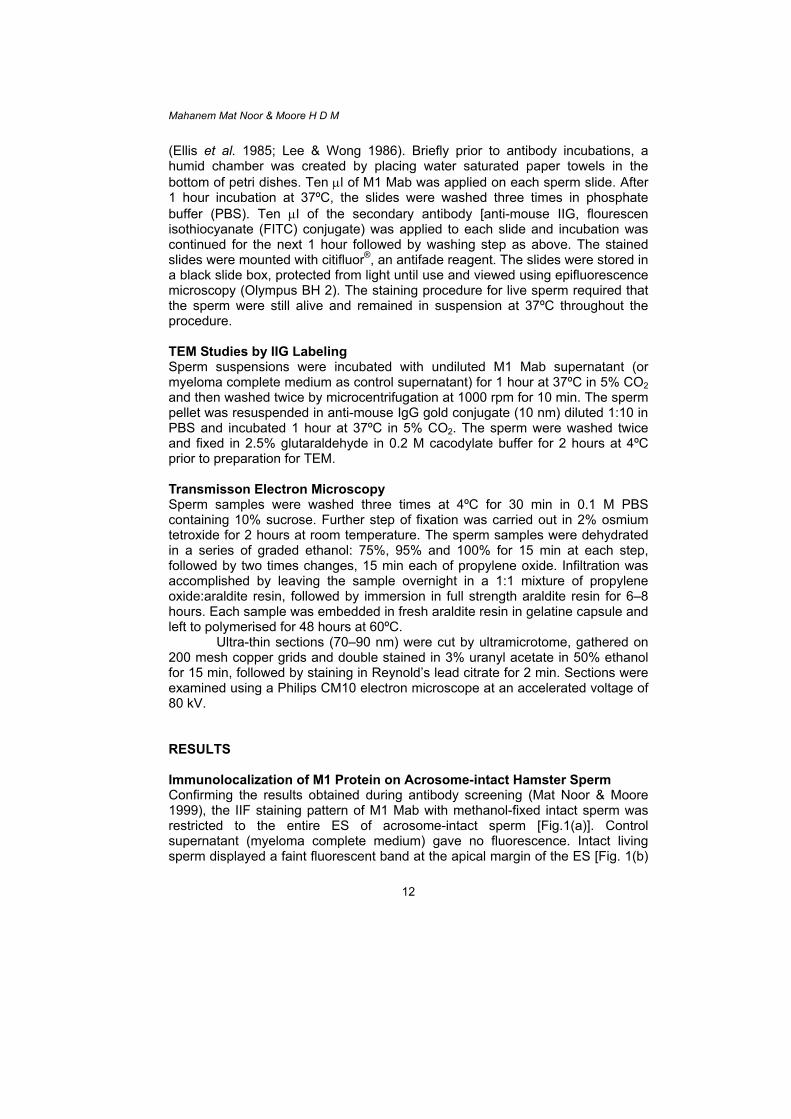
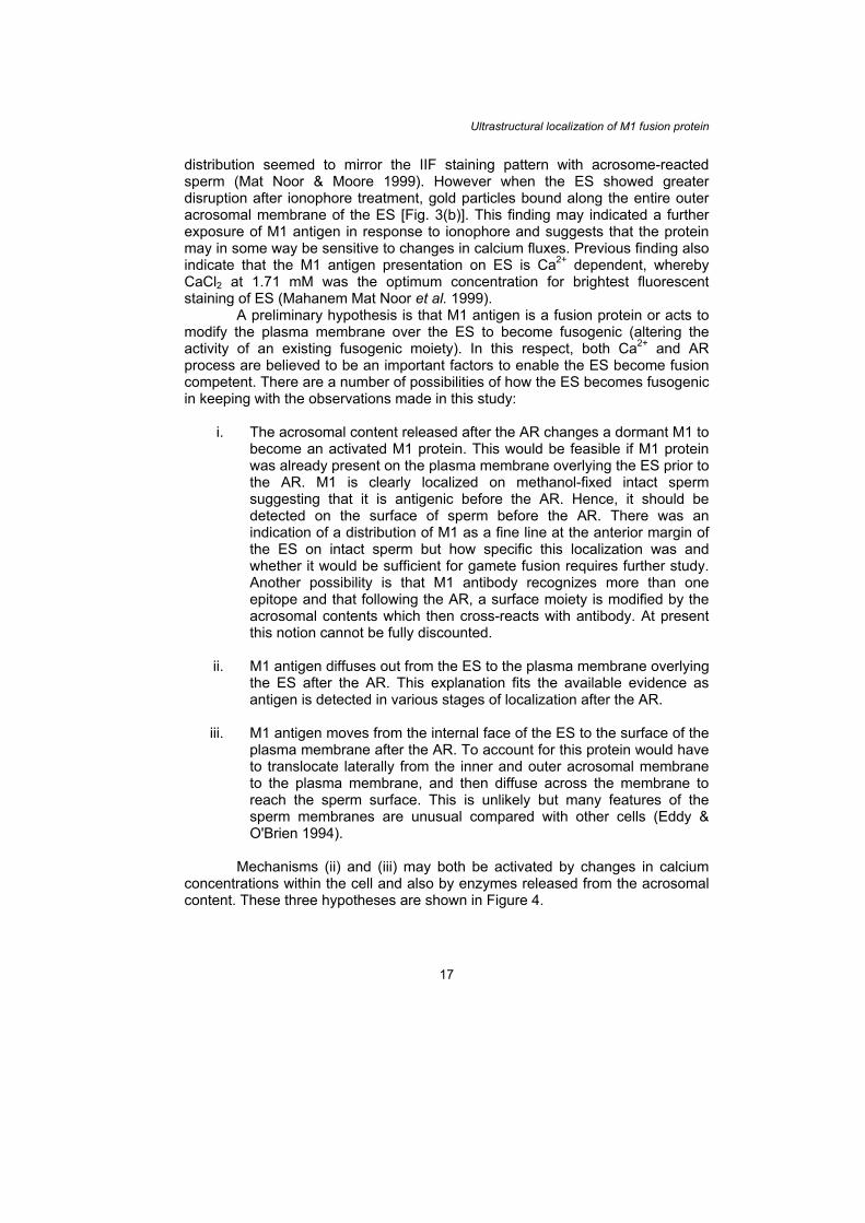
Ultra-thin sections (70–90 nm) were cut by ultramicrotome, gathered on 200 mesh copper grids and double stained in 3% uranyl acetate in 50% ethanol for 15 min, followed by staining in Reynold’s lead citrate for 2 min. Sections were examined using a Philips CM10 electron microscope at an accelerated voltage of 80 kV. RESULTS Immunolocalization of M1 Protein on Acrosome-intact Hamster Sperm Confirming the results obtained during antibody screening (Mat Noor & Moore 1999), the IIF staining pattern of M1 Mab with methanol-fixed intact sperm was restricted to the entire ES of acrosome-intact sperm [Fig.1(a)]. Control supernatant (myeloma complete medium) gave no fluorescence. Intact living sperm displayed a faint fluorescent band at the apical margin of the ES [Fig. 1(b)
12
Ultrastructural localization of M1 fusion protein & 1(b')]. In contrast, glutaraldehyde-fixed sperm (acrosome-intact) displayed no M1 localization as determined by IIG/TEM [Fig. 1(c)] when the sperm plasmalemma remained intact over the sperm head.
Figure 1: (a) IIF of intact methanol-fixed (permeabilised) hamster sperm with M1 Mab displaying fluorescent staining throughout ES region (×1000). (b) Intact living sperm displayed a faint fluorescent band at the apical margin of the ES (arrow). (b') The same sperm as (b) under normal light (×1000). (c) Electron micrograph of intact (unpermeabilised) hamster sperm after IIG with M1 Mab. No gold particles bound on sperm head (×11500).
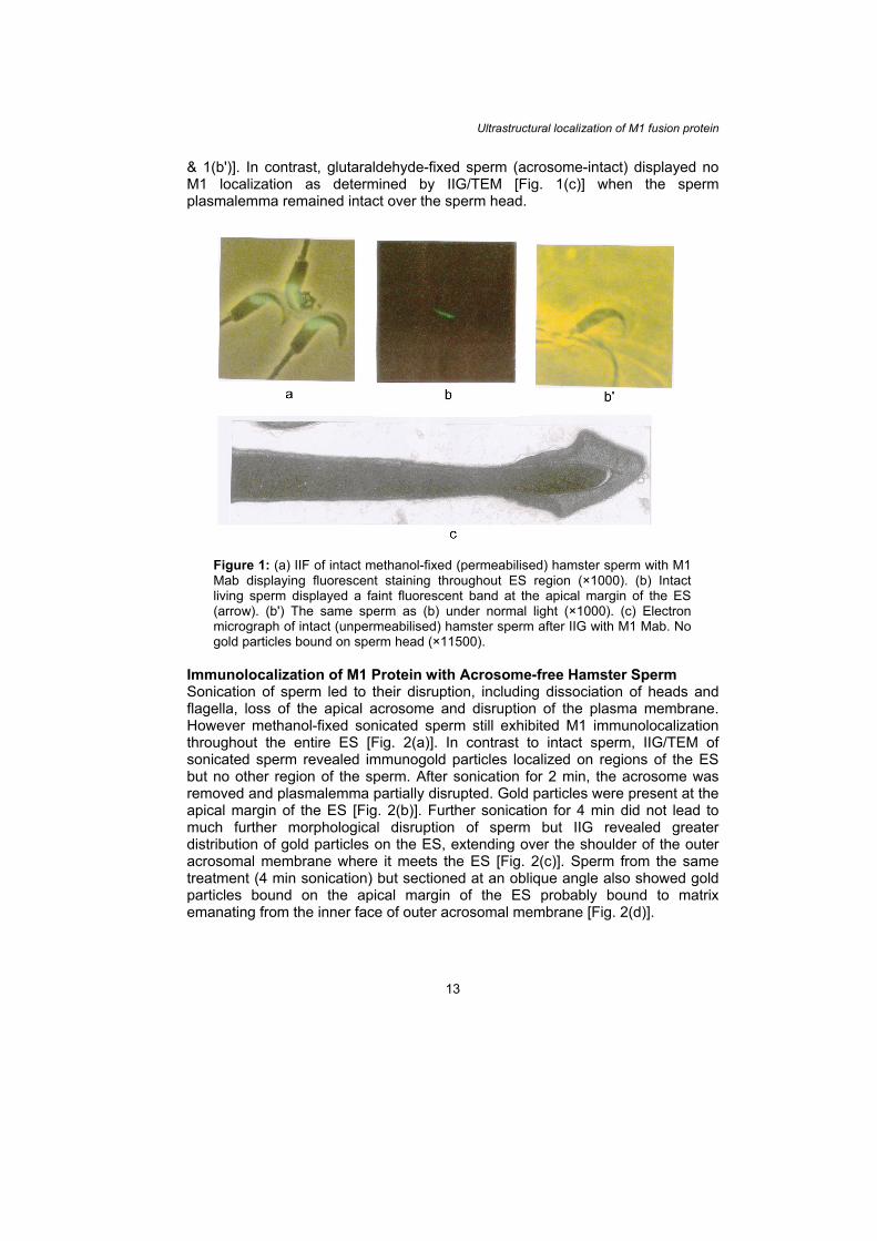
Immunolocalization of M1 Protein with Acrosome-free Hamster Sperm Sonication of sperm led to their disruption, including dissociation of heads and flagella, loss of the apical acrosome and disruption of the plasma membrane. However methanol-fixed sonicated sperm still exhibited M1 immunolocalization throughout the entire ES [Fig. 2(a)]. In contrast to intact sperm, IIG/TEM of sonicated sperm revealed immunogold particles localized on regions of the ES but no other region of the sperm. After sonication for 2 min, the acrosome was removed and plasmalemma partially disrupted. Gold particles were present at the apical margin of the ES [Fig. 2(b)]. Further sonication for 4 min did not lead to much further morphological disruption of sperm but IIG revealed greater distribution of gold particles on the ES, extending over the shoulder of the outer acrosomal membrane where it meets the ES [Fig. 2(c)]. Sperm from the same treatment (4 min sonication) but sectioned at an oblique angle also showed gold particles bound on the apical margin of the ES probably bound to matrix emanating from the inner face of outer acrosomal membrane [Fig. 2(d)].
13
Mahanem Mat Noor & Moore H D M
Figure 2: (a) IIF of sonicated methanol-fixed hamster sperm with M1 Mab showing staining throughout the ES (×400). (b)–(d) Electron micrograph of acrosome-free sperm: (b) sonicated for 2 min (×39000); (c) and (d) sonicated for 4 min at (×21000) and (×28500), respectively. All samples subjected to sequential labeling with M1 Mab and immunogold labeling. Note, gold particles consistently bound on the apical region of the ES (arrow) (b–d).
Physiological induction of the AR led to about 20% of sperm undergoing
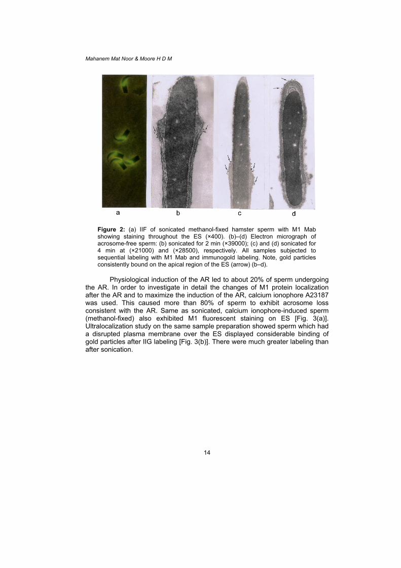
the AR. In order to investigate in detail the changes of M1 protein localization after the AR and to maximize the induction of the AR, calcium ionophore A23187 was used. This caused more than 80% of sperm to exhibit acrosome loss consistent with the AR. Same as sonicated, calcium ionophore-induced sperm (methanol-fixed) also exhibited M1 fluorescent staining on ES [Fig. 3(a)]. Ultralocalization study on the same sample preparation showed sperm which had a disrupted plasma membrane over the ES displayed considerable binding of gold particles after IIG labeling [Fig. 3(b)]. There were much greater labeling than after sonication.
14
Ultrastructural localization of M1 fusion protein
Figure 3: Acrosome-reacted hamster sperm after induction with ionophore A23187 followed by (a) IIF with M1 Mab displaying bright fluorescent staining on ES (×400). (b) Sagittal section of acrosome-reacted sperm with disruption plasma membrane displaying gold particles over outer acrosomal membrane (arrow) of ES (×46000).
DISCUSSION This study was performed to investigate the precise localization of the M1 protein on the ES of hamster sperm under different conditions. A parallel approach was employed using LM/IIF and TEM/IIG for sperm samples prepared to be either acrosome-intact or acrosome-free. As a preliminary step for immunolocalization by TEM, the effect of common fixative procedures were tested on sperm incubated with M1 Mab and evaluated by IIF. The result showed M1 Mab was not compromised with glutaraldehyde fixation because the staining became weak or lost (data not shown). Many proteins are unstable at high concentration of fixative. The cross-linking of proteins induced by aldehydes can hinder the antibody binding site by deforming the tertiary structure of the antigen (Villaroya & Scholler 1986). Some fixatives may partially or totally destroy the epitope, or else may mask antigenic sites by their insertion into surrounding glycoprotein or glycolipids (Van Ewijk et al. 1984). Hence immunolocalization at TEM level was undertaken by means of a prefixation and pre-embedding immunogold staining technique (Overstreet et al. 1995)
Methanol-fixation permeabilises the plasma membrane of sperm and therefore antibody can reach an internal antigen as well as reacting with a surface molecule. Initial studies indicated that M1 was distributed throughout the ES but not on other regions of the sperm head or tail of fixed sperm. On live
15
Mahanem Mat Noor & Moore H D M
sperm, a faint line of immunofluorescence was observed at the apical margin of the ES (Mat Noor & Moore 1999). The IIF procedure, however makes LM observations uncertain as the hamster acrosomal cap can very easily peel off and the plasma membrane can be disrupted. Therefore great care must be taken when interpreting IIF results. On the other hand, TEM reveals much more detail and the integrity of various sperm organelles can be determined with certainty. IIG labeling gives a more precise distribution of antigen but sometimes the sample preparation can lead to loss of label. The lack of immunogold particles on intact sperm suggested that M1 antigen was largely located on the interior region of the ES and was inaccessible to antibody. Occasionally, gold particles were seen on plasma membrane of intact sperm corresponding with the faint fluorescent staining seen on live intact sperm.
Although LM and TEM results indicated that M1 protein was almost exclusively an internal protein additional investigations were required to determine the exact localization. Two approaches were adopted. First, sperm samples were physically disrupted by sonication to expose the ES to the external environment and antibody. Second, sperm were induced to undergo the AR either by incubation in physiological medium alone or with the addition of calcium ionophore A23187, a commonly used and powerful inducer of the AR in mammalian sperm (Aitken et al. 1994). These different methods confirmed that M1 was an internal ES antigen but suggested that it was modified probably in response to the AR.
Physical disruption of sperm led to IIG labeling of the apical margin of the outer acrosomal membrane and the shoulder of the inner acrosomal membrane where it meets the ES. Even with prolonged sonication treatment, labeling was limited, suggesting that antigen was restricted largely to the region between the inner and outer ES with antibody access at the apical margin of the ES or to other regions of the ES when it was disrupted (Fig. 2).
Incubation of sperm in capacitating medium led to induction of the AR in a proportion of cells as determined by phase-contrast microscope observation. IIF staining revealed that M1 antigen had been exposed after the AR and had been dispersed. Compared with methanol-fixed (permeabilised) sperm, staining of live sperm was less uniform and confined usually to the more apical region of the ES, but much more intense than non-acrosome-reacted sperm. Some sperm displayed fluorescence over almost their entire ES perhaps indicating that they had undergone the AR at an earlier stage than those sperm where staining remained more marginal. The presence of staining on these cells suggested strongly that antigen had relocalized to the sperm exterior where antibody could reach (Mat Noor & Moore 1999).
To get a clear view of M1 protein localization after the AR, sperm samples were induced using the calcium ionophore A23187. In this way a large proportion of sperm could be synchronized to undergo the AR (Watson et al. 1992), facilitating TEM observations. Two distinct observations were made. Where sperm heads were displaying a characteristic AR with vesiculated apical acrosomal membrane and an intact ES (with fusion between the plasmalemma and outer acrosomal membrane) the gold particles (M1 protein) were localized over the the plasma membrane of the ES and at the apical margin of the ES. This
16
Ultrastructural localization of M1 fusion protein distribution seemed to mirror the IIF staining pattern with acrosome-reacted sperm (Mat Noor & Moore 1999). However when the ES showed greater disruption after ionophore treatment, gold particles bound along the entire outer acrosomal membrane of the ES [Fig. 3(b)]. This finding may indicated a further exposure of M1 antigen in response to ionophore and suggests that the protein may in some way be sensitive to changes in calcium fluxes. Previous finding also indicate that the M1 antigen presentation on ES is Ca2+ dependent, whereby CaCl2 at 1.71 mM was the optimum concentration for brightest fluorescent staining of ES (Mahanem Mat Noor et al. 1999).
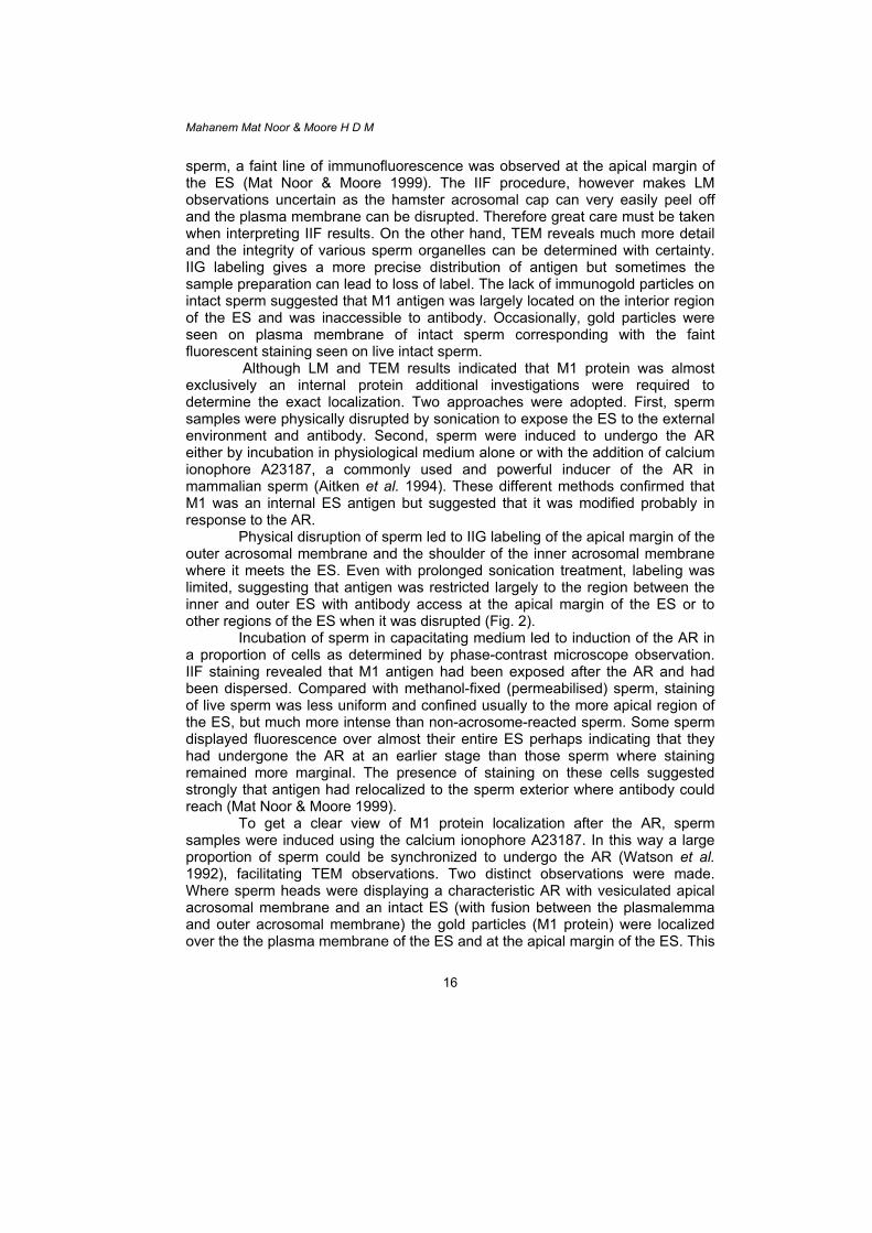
A preliminary hypothesis is that M1 antigen is a fusion protein or acts to modify the plasma membrane over the ES to become fusogenic (altering the activity of an existing fusogenic moiety). In this respect, both Ca2+ and AR process are believed to be an important factors to enable the ES become fusion competent. There are a number of possibilities of how the ES becomes fusogenic in keeping with the observations made in this study:
i. The acrosomal content released after the AR changes a dormant M1 to become an activated M1 protein. This would be feasible if M1 protein was already present on the plasma membrane overlying the ES prior to the AR. M1 is clearly localized on methanol-fixed intact sperm suggesting that it is antigenic before the AR. Hence, it should be detected on the surface of sperm before the AR. There was an indication of a distribution of M1 as a fine line at the anterior margin of the ES on intact sperm but how specific this localization was and whether it would be sufficient for gamete fusion requires further study. Another possibility is that M1 antibody recognizes more than one epitope and that following the AR, a surface moiety is modified by the acrosomal contents which then cross-reacts with antibody. At present this notion cannot be fully discounted.
ii. M1 antigen diffuses out from the ES to the plasma membrane overlying
the ES after the AR. This explanation fits the available evidence as antigen is detected in various stages of localization after the AR.
iii. M1 antigen moves from the internal face of the ES to the surface of the
plasma membrane after the AR. To account for this protein would have to translocate laterally from the inner and outer acrosomal membrane to the plasma membrane, and then diffuse across the membrane to reach the sperm surface. This is unlikely but many features of the sperm membranes are unusual compared with other cells (Eddy & O'Brien 1994).
Mechanisms (ii) and (iii) may both be activated by changes in calcium
concentrations within the cell and also by enzymes released from the acrosomal content. These three hypotheses are shown in Figure 4.
17
Mahanem Mat Noor & Moore H D M
b
Figure 4: The hypothesis of the M1 protein movements during or after AR toenables ES becomes fusogenic: (a) sperm before AR, (b) sperm during or afteAR and (c) fusogenic sperm.
The localization of M1 protein as investigated in this study rema
consistent with a possible role in gamete fusion. The restricted binding oantibody to the ES was similar to that of monoclonal antibodies G11 and reported to bind to the ES of guinea pig sperm (Allen & Green 1995)monoclonal antibody M29 which was shown to bind to the ES of mouse sp(Saling et al. 1985) and MN9 who was reported as the monoclonal antibodyrecognized fusion protein on ES of mouse sperm (Toshimori et al. 1998).
In conclusion, the results obtained in this study clearly indicate that tis a very close relationship between the AR, Ca2+ fluxes with M1 anpresentation and/or activation on ES hamster sperm. Further informconcerning the M1 antigen is required before it could be ascertained exactlythe antigen might initiate sperm-egg fusion or its role in fertilization processes ACKNOWLEDGEMENTS Financial assistance from the Malaysian Government and University of ShefUnited Kingdom are acknowledged.
18
IAM
a cr
ined f M1 M13, , the erm
that
here tigen ation how .
field,
Ultrastructural localization of M1 fusion protein REFERENCES Aitken J, Buckingham D and Harkiss D. (1994). Analysis of the extent to which sperm
movement can predict the results of ionophore-enhanced functional assays of the acrosome reaction and sperm-oocyte fusion. Human Reprod. 9(10): 1867–1874.
Allen C A and Green D P L. (1995). Monoclonal antibodies which recognize equatorial
segment epitopes presented de novo following the A23187-induced acrosome reaction of guinea pig sperm. J. Cell Science 108: 767–777.
Barros C and Herrera E. (1977). Ultrastructural observations of the incorporation of guinea
pig spermatozoa into zona free hamster oocytes. J. Reprod. Fertil. 49: 47–50. Bedford J M, Moore H D M and Franklin L E. (1979). Significance of the equatorial
segment of the acrosome of the spermatozoa in eutherian mammals. Exp. Cell. Res. 119: 119–126.
Biggers J D, Whitten W K and Whittingham D G. (1971). The culture of mouse embryos in
vitro. In: Daniel J C (ed.). Methods in mammalian embryology. San Francisco: Freeman W H and Company, pp. 86–116.
Bronson R A and Fusi F M. (1996). Integrins and human reproduction. Mol. Hum. Reprod.
2(3): 153–168. Eddy E M and O'Brien D A. (1994). The spermatozoon. In: Knobil E and Neil J D (eds.).
The physiology of reproduction. New York: Raven Press, pp. 29–77. Ellis D H, Hartman T D and Moore H D M. (1985). Maturation and function of the hamster
spermatozoon probed with monoclonal antibodies. J. Reprod. Immunol. 7: 299–314.
Kohler J K, de Curtis I, Stenchever M A and Smith D. (1982). Interaction of human sperm
with zona-free hamster eggs: A freeze-fracture study. Gamete Res. 6: 371–386. Lee C Y G and Wong E. (1986). Developmental studies of sperm surface antigens using
sperm-specific monoclonal antibodies. J. Reprod. Immunol. 9: 275–287. Mat Noor M and Moore H D M. (1999). Monoclonal antibody that recognizes an epitope of
the sperm equatorial region and specifically inhibits sperm-oolemma fusion but not binding. J. Reprod. Fertil. 115: 215–224.
Mahanem Mat Noor, Jumaidah M G and Moore H D M. (1999). Correlation of Ca2+ ion and
acrosome reaction with presentation of M1 fusion protein in hamster spermatozoa. Malay. Appl. Biol. 28: 119–124.
Moore H D M and Bedford J M. (1978). Ultrastructure of the equatorial segment of
hamster spermatozoa during penetration of oocytes. J. Ultrastruct. Res. 62: 110–117.
Overstreet J W, Lin Y, Yudin A I, Meyers S A, Primakoff P, Myles D G, Katz D F and
Vandevoort C A. (1995). Location of the PH-20 protein in acrosome-intact and
19
Mahanem Mat Noor & Moore H D M
acrosome-reacted spermatozoa of Cynomolgus macaques. Biol. Reprod. 52: 105–114.
Saling P M, Irons G and Waibel R. (1985). Mouse sperm antigens that participate in
fertilization. I. Inhibition of sperm fusion with the egg plasma membrane using monoclonal antibodies. Biol. Reprod. 33: 515–526.
Sathananthan A H, Ng S C, Edirisinghe R, Ratnam S S and Wong P C. (1986). Sperm-
oocyte interaction in the human during polyspermic fertilization in vitro. Gamete Res. 15: 317–326.
Taggart D A, O'ien H and Moore H D M. (1993). Ultrastructural characteristics of in vitro
fertilization in the grey short-tailed opossum, Monodelphis domestica. Anat. Rec. 237: 21–37.
Toshimori K, Saxena D K, Tanii I and Yoshinaga K. (1998). An MN9 antigenic molecule,
equatorin, is required for successful sperm-oocyte fusion in mice. Biol. Reprod. 59: 22–29.
Van Ewijk W, Van Soest P L, Verkerk A and Jongkind J F. (1984). Loss of antibody to
prefixed cells: Fixation parameters for immunocytochemistry. Histochem. J. 16: 179–193.
Villarroya S and Scholler R. (1986). Regional heterogeneity of human spermatozoa
detected with monoclonal antibodies. J. Reprod. Fertil. 76: 435–447. Watson P F, Plummer J M and Jones P S. (1992). The ionophore-induced acrosome
reaction differs structurally from the spontaneous acrosome reaction. J. Exp. Zool. 264(2): 231–235.
Yanagimachi R. (1994). Mammalian fertilization. In: Knobil E and Neil J D (eds.). The
physiology of reproduction. 2nd ed. New York: Raven Press, 1: 189–317.
20


![[M1] Percepatan Gravitasi Bumi](https://static.fdokumen.site/doc/165x107/55cf977d550346d03391e827/m1-percepatan-gravitasi-bumi.jpg)